 Explore
 Validate
 Learn
Learn Western blot
Antibody data
- Antibody Data
- Antigen structure
- References [0]
- Comments [0]
- Validations
- Western blot [3]
- Immunocytochemistry [1]
- Immunohistochemistry [1]
Submit
Validation data
Reference
Comment
Report error
- Product number
- PA5-18022 - Provider product page

- Provider
- Invitrogen Antibodies
- Product name
- NQO1 Polyclonal Antibody
- Antibody type
- Polyclonal
- Antigen
- Synthetic peptide
- Description
- This antibody is predicted to react with porcine and rat based on sequence homology. This antibody is tested in Peptide ELISA: antibody detection limit dilution 64,000.
- Reactivity
- Human, Rat
- Host
- Goat
- Isotype
- IgG
- Vial size
- 100 µg
- Concentration
- 0.5 mg/mL
- Storage
- -20° C, Avoid Freeze/Thaw Cycles
No comments: Submit comment
Supportive validation
- Submitted by
- Invitrogen Antibodies (provider)
- Main image

- Experimental details
- Western blot analysis of NQO1 in Rat (A) and Pig (B) Kidney lysate (35µg protein in RIPA buffer). Samples were probed with the NQO1 antibody (Product # PA5-18022, 1µg/mL) for 1 hour. Western blot was detected by chemiluminescence.

- Submitted by
- Invitrogen Antibodies (provider)
- Main image

- Experimental details
- Western Blot staining of Human Kidney lysate using Product # PA5-18022 at a concentration of 0.03 µg/mL, the primary antibody incubation was 1 hour and the detection method was chemiluminescence.

- Submitted by
- Invitrogen Antibodies (provider)
- Main image

- Experimental details
- Western blot analysis was performed on whole cell extracts (30 µg lysate) of Hep G2 (Lane 1), HeLa (Lane 2), A549 (Lane 3), U-87 MG (Lane 4), SH-SY5Y (Lane 5) and MOLT4 (Lane 6). The blot was probed with Anti-NQO1 Polyclonal Antibody (Product # PA5-18022, 1:1000 dilution) and detected by chemiluminescence using Rabbit anti-Goat IgG (H+L) Superclonal™ Secondary Antibody, HRP conjugate (Product # A27014, 0.25 µg/ml, 1:4000 dilution). A 32 kDa band corresponding to NQO1 was detected across the cell lines tested except SH-SY5Y and MOLT4 which showed little or no expression respectively as reported in the literature. An additional non-specific band (*) was observed at ~70 kDa.

Supportive validation
- Submitted by
- Invitrogen Antibodies (provider)
- Main image
- Experimental details
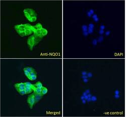
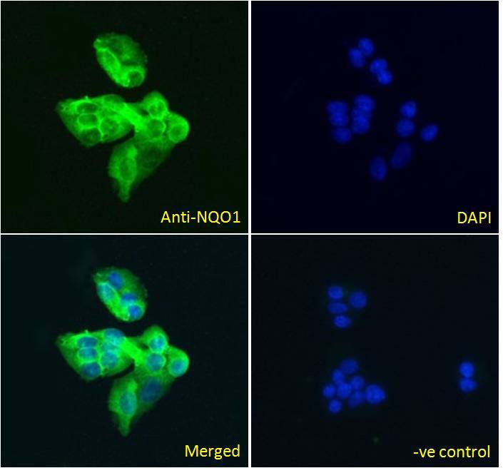
- Immunofluorescence analysis of NQO1 in HepG2 cells using a NQO1 monoclonal antibody (Product # PA5-18022) at 5 µg/mL for1hr. The cells were paraformaldehyde fixed and permeabilized with 0.15% Triton. Primary incubation was followed by Alexa Fluor 488 secondary antibody (2 µg/mL) showing cytoplasmic staining. The nuclear stain is DAPI (blue). Negative control: Unimmunized goat IgG (5 µg/mL)followed by Alexa Fluor 488 secondary antibody (2 µg/mL).
Supportive validation
- Submitted by
- Invitrogen Antibodies (provider)
- Main image

- Experimental details
- Immunohistochemical analysis of NQO1 in paraffin embedded human kidney using a NQO1 polyclonal antibody (Product #PA5-18022) at a concentration of 5 µg/mL. Steamed antigen retrieval was performed with pH 6 buffered citrate. Samples were then stained with alkaline phosphatase.
