Explore
 Validate
 Learn
LearnPA5-22288
antibody from Invitrogen Antibodies
Targeting: PSMA6
IOTA, MGC22756, MGC2333, MGC23846, p27K, PROS27
 Western blot
 Immunocytochemistry
Antibody data
- Antibody Data
- Antigen structure
- References [1]
- Comments [0]
- Validations
- Immunocytochemistry [1]
- Immunohistochemistry [4]
- Other assay [2]
Submit
Validation data
Reference
Comment
Report error
- Product number
- PA5-22288 - Provider product page

- Provider
- Invitrogen Antibodies
- Product name
- PSMA6 Polyclonal Antibody
- Antibody type
- Polyclonal
- Antigen
- Recombinant full-length protein
- Description
- Recommended positive controls: 293T, A431, H1299, HeLa, mouse liver, rat liver. Predicted reactivity: Mouse (100%), Rat (100%), Xenopus laevis (87%), Pig (98%), Chicken (96%), Sheep (99%), Rhesus Monkey (100%), Chimpanzee (100%), Bovine (100%). Store product as a concentrated solution. Centrifuge briefly prior to opening the vial.
- Reactivity
- Human, Mouse, Rat
- Host
- Rabbit
- Isotype
- IgG
- Vial size
- 100 μL
- Concentration
- 1 mg/mL
- Storage
- Store at 4°C short term. For long term storage, store at -20°C, avoiding freeze/thaw cycles.
Submitted references Low Neurotoxicity of ONX-0914 Supports the Idea of Specific Immunoproteasome Inhibition as a Side-Effect-Limiting, Therapeutic Strategy.
von Brzezinski L, Säring P, Landgraf P, Cammann C, Seifert U, Dieterich DC
European journal of microbiology & immunology 2017 Sep;7(3):234-245
European journal of microbiology & immunology 2017 Sep;7(3):234-245
No comments: Submit comment
Supportive validation
- Submitted by
- Invitrogen Antibodies (provider)
- Main image

- Experimental details
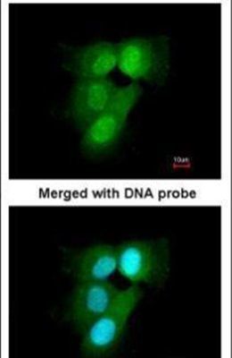
- Immunofluorescent analysis of 20S Proteasome alpha6 in paraformaldehyde-fixed A431 cells using a 20S Proteasome alpha6 polyclonal antibody (Product # PA5-22288) at a 1:200 dilution.
Supportive validation
- Submitted by
- Invitrogen Antibodies (provider)
- Main image
- Experimental details
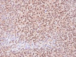
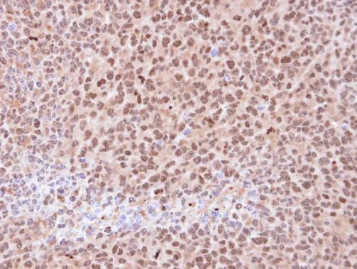
- Immunohistochemical analysis of paraffin-embedded H1299 xenograft , using 20S Proteasome alpha6 (Product # PA5-22288) antibody at 1:100 dilution. Antigen Retrieval: EDTA based buffer, pH 8.0, 15 min.
- Submitted by
- Invitrogen Antibodies (provider)
- Main image
- Experimental details
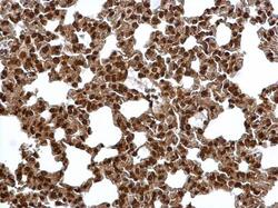
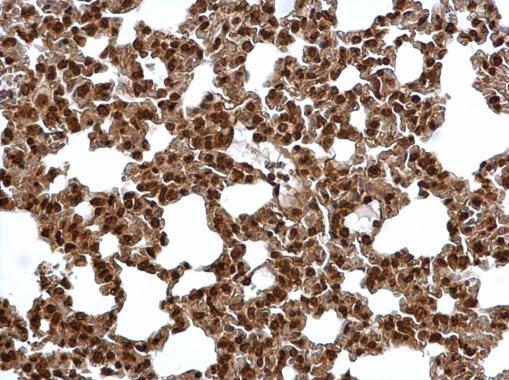
- Proteasome 20S alpha 6 antibody detects Proteasome 20S alpha 6 protein at cytosol and nucleus on mouse lung by immunohistochemical analysis. Sample: Paraffin-embedded mouse lung. Proteasome 20S alpha 6 antibody (Product # PA5-22288) dilution: 1:500. Antigen Retrieval: EDTA based buffer, pH 8.0, 15 min.
- Submitted by
- Invitrogen Antibodies (provider)
- Main image
- Experimental details
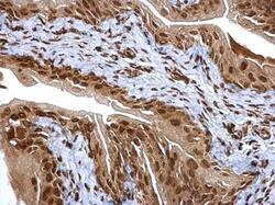
- Proteasome 20S alpha 6 antibody detects Proteasome 20S alpha 6 protein at cytosol and nucleus on mouse urinary bladder by immunohistochemical analysis. Sample: Paraffin-embedded mouse urinary bladder. Proteasome 20S alpha 6 antibody (Product # PA5-22288) dilution: 1:500. Antigen Retrieval: EDTA based buffer, pH 8.0, 15 min.

- Submitted by
- Invitrogen Antibodies (provider)
- Main image

- Experimental details
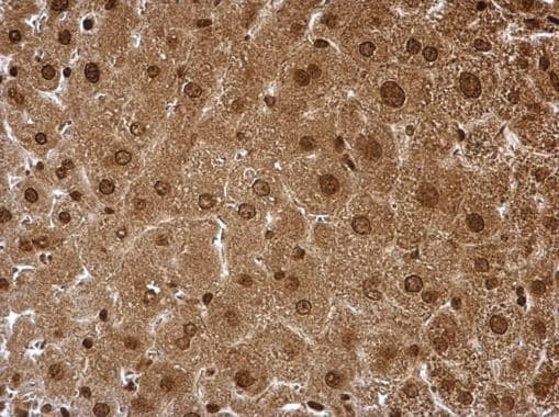
- Proteasome 20S alpha 6 antibody detects Proteasome 20S alpha 6 protein at cytosol and nucleus on mouse liver by immunohistochemical analysis. Sample: Paraffin-embedded mouse liver. Proteasome 20S alpha 6 antibody (Product # PA5-22288) dilution: 1:500. Antigen Retrieval: EDTA based buffer, pH 8.0, 15 min.
Supportive validation
- Submitted by
- Invitrogen Antibodies (provider)
- Main image

- Experimental details
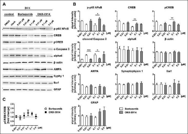
- Fig. 4. Bortezomib and ONX-0914 differentially affect neural protein expression after 24 h. Cell lysates of primary cortical cultures (DIV14), treated for 24 h with indicated inhibitor concentrations, were analysed via immunoblot for selected proteins (A) and signal intensities were subsequently determined (B). Normalization was ensured via loading same protein amounts and quantitative analysis of the respective Coomassie stained sister gel. Data were expressed in relation to the control condition (100%). Note, protein levels for NFkappaB, CREB and pCREB showed a tendency towards decreased expression with increasing inhibitor concentrations, whereas this effect was evident earlier and stronger on Bortezomib treatment compared to ONX-0914. Conversely, levels of the apoptosis marker Caspase 3 were significantly increased after Bortezomib treatment at doses of 0.01 and 0.1 uM after 24 h. No significant differences were detected for the protein level of alpha6, beta-actin, AMPA and Synaptophysin 1 as well as the glial marker Iba1 and GFAP after 24 h inhibitor treatment (4B). Activation of CREB signalling was assessed by calculation of the pCREB/CREB ratio (4C). A slight tendency for CREB activation indicated by phosphorylation was observed after 24 h at doses of 0.1 uM for both inhibitors. Presented data are mean +-SEM of 2-3 technical replicates each from 3 independent experiments, B: Student's t -test, *: p < 0.05, **: p < 0.01, ***: p < 0.001, ****: p < 0.0001, C: one-sample t
- Submitted by
- Invitrogen Antibodies (provider)
- Main image
- Experimental details
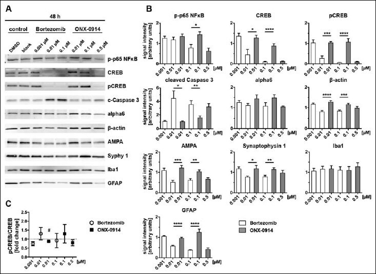
- Fig. 5. Altered neural protein expression after 48 h of inhibitor treatment. Alterations of protein levels after proteasome inhibition were detected by immunoblot analysis (A) and the signal intensities subsequently quantified (B). Cell lysates were prepared from primary cortical co-cultures after 48 h inhibitor treatment. Identical protein concentrations were loaded on gels and signals normalized to Coomassie stained sister gels. Signal intensities are expressed in relation to the control (100%). After 48 h, effects on protein expression after inhibitor treatment became even more obvious for NFkappaB and Caspase 3. The prolonged inhibitor incubation at a dose of 0.01 uM additionally effected massively decreased levels for CREB and pCREB after 48 h Bortezomib application compared to ONX-0914. Further, treatment with 0.01 and 0.1 uM Bortezomib over 48 h lead to decreased protein levels for beta-actin, AMPA, Synaptophysin 1 and GFAP in comparison to ONX-0914-treated cultures. Expression levels for alpha6 subunit and Iba1 were unaffected by inhibitor treatment over 48 h (5B). CREB activation indicated by phosphorylation was, compared to 24 h (4C), nearly unaffected after 48 h (5C). Presented data are mean +-SEM of 2-3 technical replicates each from 3 independent experiments, B: Student's t -test, *: p < 0.05, **: p < 0.01, ***: p < 0.001, ****: p < 0.0001, C: one-sample t -test against hypothetical value 1, # : p < 0.05