Explore
 Validate
 Learn
Learn Western blot
 Other assay
Antibody data
- Antibody Data
- Antigen structure
- References [1]
- Comments [0]
- Validations
- Other assay [2]
Submit
Validation data
Reference
Comment
Report error
- Product number
- PA5-103350 - Provider product page

- Provider
- Invitrogen Antibodies
- Product name
- ALPK3 Polyclonal Antibody
- Antibody type
- Polyclonal
- Antigen
- Synthetic peptide
- Description
- Antibody detects endogenous levels of total ALPK3.
- Reactivity
- Human, Mouse
- Host
- Rabbit
- Isotype
- IgG
- Vial size
- 100 μL
- Concentration
- 1 mg/mL
- Storage
- -20°C
Submitted references MicroRNA-384-5p protects against cardiac hypertrophy via the ALPK3 signaling pathway.
Guo S, Yang Y, Qian W, Yao Y, Zhou G, Shen L, Zhou J
Journal of biochemical and molecular toxicology 2022 Aug;36(8):e23093
Journal of biochemical and molecular toxicology 2022 Aug;36(8):e23093
No comments: Submit comment
Supportive validation
- Submitted by
- Invitrogen Antibodies (provider)
- Main image

- Experimental details
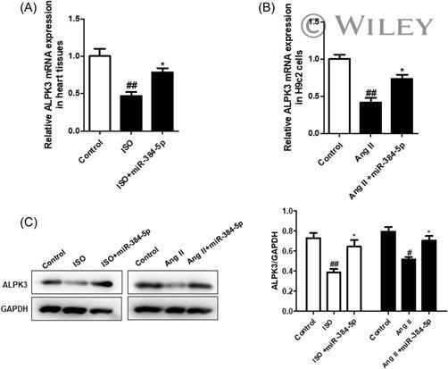
- 4 Figure miR-384-5p regulates the expression of ALPK3 in the progression of cardiac hypertrophy. The ALPK3 mRNA expression levels in mice cardiac tissue (A) and H9c2 (B) in the cardiac hypertrophy model were measured by quantitative reverse transcription polymerase chain reaction. (C) The protein expression of ALPK3 was measured using western blot. Data are presented as the mean +- standard error of the mean ( n = 3). # p < 0.05, ## p < 0.01 compared with the control group; * p < 0.05, ** p < 0.01 compared with the isoproterenol group or Ang-II group.
- Submitted by
- Invitrogen Antibodies (provider)
- Main image

- Experimental details
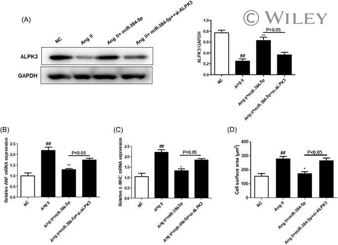
- 5 Figure The low expression of ALPK3 can aggravate the myocardial hypertrophy inhibitory activity of miR-384-5p. Primary cultures of cardiac myocytes were treated with Ang-II (1 mM) for 48 h and cotransfected with miR-384-5p mimics and the empty vector or plasmid of the ALPK3 gene without the 3'-UTR. (A) Western blot analysis of ALPK3 protein expression. (B) The ANF and beta-MHC mRNA expression levels were measured using quantitative reverse transcription polymerase chain reaction. (E) The surface areas of cardiomyocytes were quantified. Data are presented as the mean +- standard error of the mean ( n = 3). # p < 0.05, ## p < 0.01 compared with the NC + empty vector group. * p < 0.05, ** p < 0.01 compared with the miR-384-5p + Vector group. NC, negative control; UTR, untranslated region.