 Explore
 Validate
 Learn
Learn Western blot
 Immunocytochemistry
Antibody data
- Antibody Data
- Antigen structure
- References [1]
- Comments [0]
- Validations
- Western blot [1]
- Immunocytochemistry [1]
- Immunohistochemistry [1]
Submit
Validation data
Reference
Comment
Report error
- Product number
- AMAb90598 - Provider product page

- Provider
- Atlas Antibodies
- Proper citation
- Atlas Antibodies Cat#AMAb90598, RRID:AB_2665602
- Product name
- Anti-S100A4
- Antibody type
- Monoclonal
- Description
- Monoclonal Antibody against Human S100A4, Clone ID: CL0239, Gene description: S100 calcium binding protein A4, Alternative Gene Names: 18A2, 42A, CAPL, FSP1, MTS1, P9KA, PEL98, Validated applications: ICC, IHC, WB, Uniprot ID: P26447, Storage: Store at +4°C for short term storage. Long time storage is recommended at -20°C.
- Reactivity
- Human
- Host
- Mouse
- Conjugate
- Unconjugated
- Isotype
- IgG
- Antibody clone number
- CL0239
- Vial size
- 100 µl
- Concentration
- 0.4 mg/ml
- Storage
- Store at +4°C for short term storage. Long time storage is recommended at -20°C.
- Handling
- The antibody solution should be gently mixed before use.
Submitted references The drug efficacy testing in 3D cultures platform identifies effective drugs for ovarian cancer patients
Åkerlund E, Gudoityte G, Moussaud-Lamodière E, Lind O, Bwanika H, Lehti K, Salehi S, Carlson J, Wallin E, Fernebro J, Östling P, Kallioniemi O, Joneborg U, Seashore-Ludlow B
npj Precision Oncology 2023;7(1)
npj Precision Oncology 2023;7(1)
No comments: Submit comment
Enhanced validation
- Submitted by
- Atlas Antibodies (provider)
- Enhanced method
- Genetic validation
- Main image

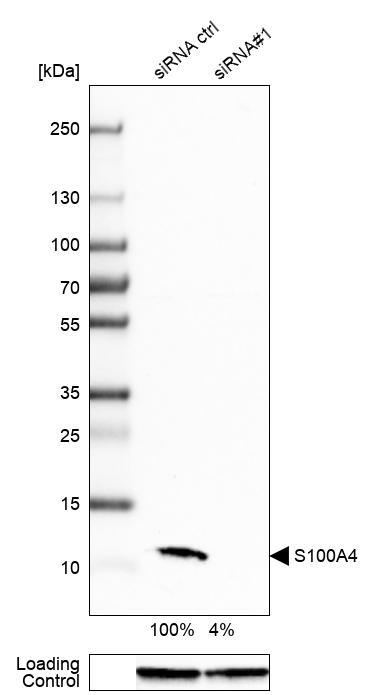
- Experimental details
- Western blot analysis in A-549 cells transfected with control siRNA, target specific siRNA probe #1, using Anti-S100A4 antibody. Remaining relative intensity is presented. Loading control: Anti-GAPDH.
- Sample type
- Human
- Protocol
- Protocol
Supportive validation
- Submitted by
- Atlas Antibodies (provider)
- Main image

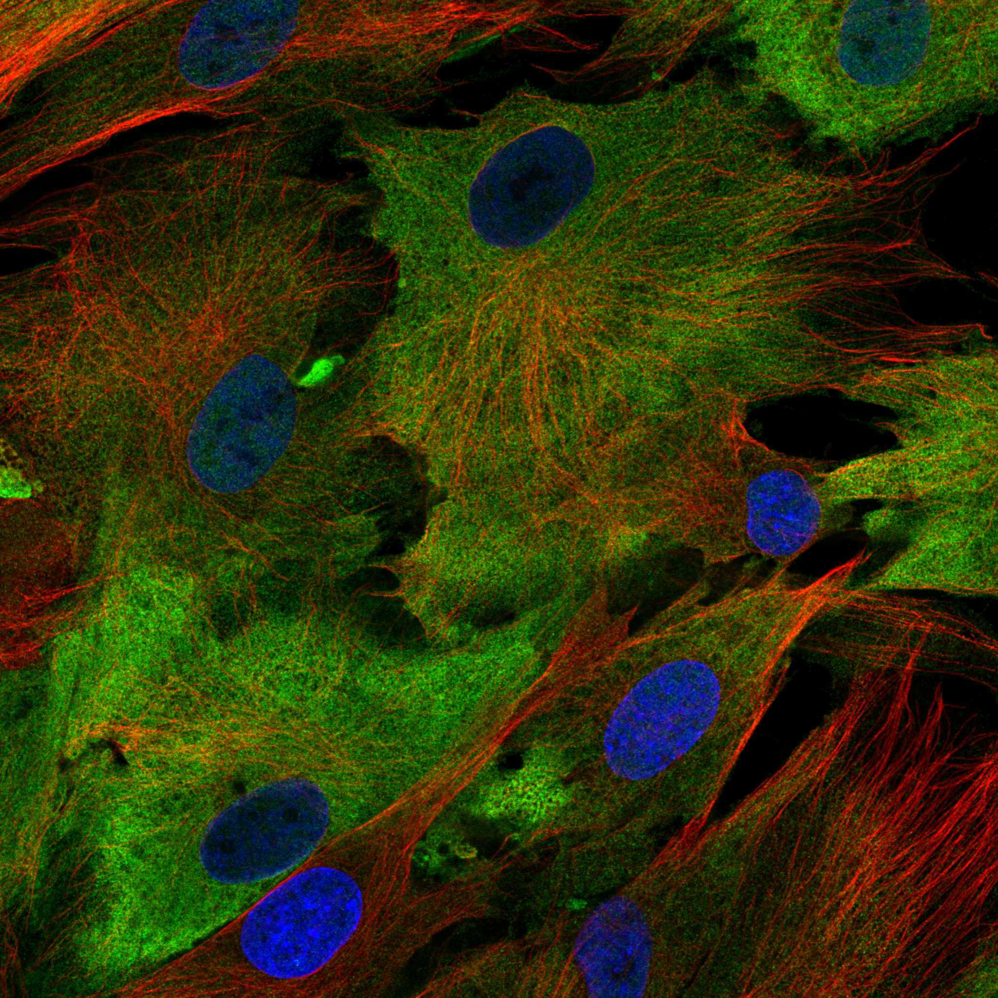
- Experimental details
- Immunofluorescence staining of BJ cells using the anti-S100A4 monoclonal antibody, showing specific staining in the plasma membrane in green. Microtubule- and nuclear probes are visualized in red and blue, respectively (where available).
- Sample type
- Human
Supportive validation
- Submitted by
- Atlas Antibodies (provider)
- Enhanced method
- Orthogonal validation
- Main image
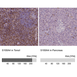
- Experimental details
- Immunohistochemistry analysis in human tonsil and pancreas tissues using AMAb90598 antibody. Corresponding S100A4 RNA-seq data are presented for the same tissues.
- Sample type
- Human
- Protocol
- Protocol
