 Explore
 Validate
 Learn
LearnNBP2-36431
antibody from Novus Biologicals
Targeting: S100A4
18A2, 42A, CAPL, FSP1, MTS1, P9KA, PEL98
 Western blot
 Immunocytochemistry
Antibody data
- Antibody Data
- Antigen structure
- References [0]
- Comments [0]
- Validations
- Western blot [2]
- Immunohistochemistry [5]
- Flow cytometry [2]
Submit
Validation data
Reference
Comment
Report error
- Product number
- NBP2-36431 - Provider product page

- Provider
- Novus Biologicals
- Product name
- Mouse Monoclonal S100A4 Antibody
- Antibody type
- Monoclonal
- Description
- Protein G purified.
- Reactivity
- Human
- Host
- Mouse
- Isotype
- IgG
- Vial size
- 0.1 mg
- Concentration
- 1.0 mg/ml
- Storage
- Store at 4C short term. Aliquot and store at -20C long term. Avoid freeze-thaw cycles.
No comments: Submit comment
Supportive validation
- Submitted by
- Novus Biologicals (provider)
- Main image
- Experimental details
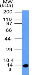
- Western Blot: S100A4 Antibody (1C4) [NBP2-36431] - WB analysis of S100A4 recombinant protein with S100A4 antibody (clone 1C4) at 0.5 ug/ml concentration.

- Submitted by
- Novus Biologicals (provider)
- Main image

- Experimental details
- Western Blot: S100A4 Antibody (1C4) [NBP2-36431] - WB analysis of S100A4 in lysates of A375 and A549 cell lines with S100A4 antibody (clone 1C4). The antibody detected the endogenous protein at its expected molecular weight position 11.7 kDa.

Supportive validation
- Submitted by
- Novus Biologicals (provider)
- Main image
- Experimental details
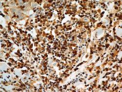
- Immunohistochemistry-Paraffin: S100A4 Antibody (1C4) [NBP2-36431] - Aanalysis of tissue section of Squamous Cell Carcinoma of Human Lungs with mouse monoclonal S100A4 antibody (clone 1C4) at 5 ug/ml concentration. An expected cytoplasmic as well as nuclear staining was observed in the tissue and the staining was found to be more intense in several cells which are potentially the advanced cancer cells, lymphocytes or infiltrating macrophages.

- Submitted by
- Novus Biologicals (provider)
- Main image

- Experimental details
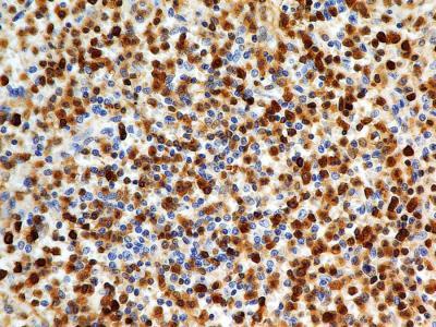
- Immunohistochemistry-Paraffin: S100A4 Antibody (1C4) [NBP2-36431] - Analysis of tissue section of normal human spleen with mouse monoclonal S100A4 antibody (clone 1C4) at 5 ug/ml concentration.
- Submitted by
- Novus Biologicals (provider)
- Main image
- Experimental details
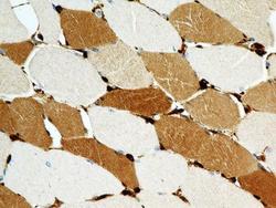
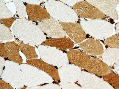
- Immunohistochemistry-Paraffin: S100A4 Antibody (1C4) [NBP2-36431] - Analysis of tissue section of normal human skeletal muscle with mouse monoclonal S100A4 antibody (clone 1C4) at 5 ug/ml concentration. The different myocytes showed a varying degree of cytoplasmic immuno-positivity whereas the staining was found to be intense in some of the peripherally placed nuclei.
- Submitted by
- Novus Biologicals (provider)
- Main image

- Experimental details
- Immunohistochemistry-Paraffin: S100A4 Antibody (1C4) [NBP2-36431] - Analysis of tissue section of human normal lung with mouse monoclonal S100A4 antibody (clone 1C4) at 5 ug/ml concentration. Majority of the cells of respiratory alveoli showed the cytoplasmic staining whereas some clusters of cells (lymphocytes, macrophages) developed nuclear staining also.

- Submitted by
- Novus Biologicals (provider)
- Main image

- Experimental details
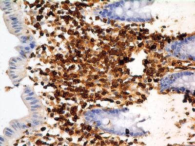
- Immunohistochemistry-Paraffin: S100A4 Antibody (1C4) [NBP2-36431] - Analysis of tissue section of human colon with mouse monoclonal S100A4 antibody (clone 1C4) at 5 ug/ml concentration. The columnar epithelial cells showed weak to negligible immunopositivity whereas the cells present in lamina propria, the loose cellular connective tissue in mucosa, developed intense staining in various type of cells.
Supportive validation
- Submitted by
- Novus Biologicals (provider)
- Main image

- Experimental details
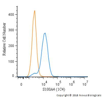
- Flow Cytometry: S100A4 Antibody (1C4) [NBP2-36431] - An intracellular stain was performed on HeLa cells with S100A4 (1C4) Antibody NBP2-36431 and a matched isotype control. Cells were fixed with 4% PFA and then permeabilized with 0.1% saponin. Cells were incubated in an antibody dilution of 5.0 ug/mL for 30 minutes at room temperature, followed by Rabbit IgG (H+L) Cross-Adsorbed Secondary Antibody, Dylight 550.
- Submitted by
- Novus Biologicals (provider)
- Main image
- Experimental details
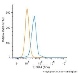
- Flow Cytometry: S100A4 Antibody (1C4) [NBP2-36431] - An intracellular stain was performed on THP-1 cells with S100A4 (1C4) Antibody NBP2-36431 and a matched isotype control. Cells were fixed with 4% PFA and then permeabilized with 0.1% saponin. Cells were incubated in an antibody dilution of 5.0 ug/mL for 30 minutes at room temperature, followed by Rabbit IgG (H+L) Cross-Adsorbed Secondary Antibody, Dylight 550.
