Explore
 Validate
 Learn
LearnMA5-32347
antibody from Invitrogen Antibodies
Targeting: S100A4
18A2, 42A, CAPL, FSP1, MTS1, P9KA, PEL98
 Western blot
 Immunocytochemistry
Antibody data
- Antibody Data
- Antigen structure
- References [2]
- Comments [0]
- Validations
- Immunocytochemistry [6]
- Immunohistochemistry [7]
- Flow cytometry [2]
- Other assay [1]
Submit
Validation data
Reference
Comment
Report error
- Product number
- MA5-32347 - Provider product page

- Provider
- Invitrogen Antibodies
- Product name
- S100A4 Recombinant Rabbit Monoclonal Antibody (SD200-08)
- Antibody type
- Monoclonal
- Antigen
- Recombinant full-length protein
- Description
- Recombinant rabbit monoclonal antibodies are produced using in vitro expression systems. The expression systems are developed by cloning in the specific antibody DNA sequences from immunoreactive rabbits. Then, individual clones are screened to select the best candidates for production. The advantages of using recombinant rabbit monoclonal antibodies include: better specificity and sensitivity, lot-to-lot consistency, animal origin-free formulations, and broader immunoreactivity to diverse targets due to larger rabbit immune repertoire.
- Reactivity
- Human, Mouse, Rat
- Host
- Rabbit
- Isotype
- IgG
- Antibody clone number
- SD200-08
- Vial size
- 100 μL
- Concentration
- 1 mg/mL
- Storage
- Store at 4°C short term. For long term storage, store at -20°C, avoiding freeze/thaw cycles.
Submitted references A conserved pathway of transdifferentiation in murine Kupffer cells.
PECAM1 Combines With CXCR4 to Trigger Inflammatory Cell Infiltration and Pulpitis Progression Through Activating the NF-κB Signaling Pathway.
Li X, Hollingshead N, Lampert S, Truong CD, Li W, Niu J, Crispe IN, Soysa R
European journal of immunology 2021 Oct;51(10):2452-2463
European journal of immunology 2021 Oct;51(10):2452-2463
PECAM1 Combines With CXCR4 to Trigger Inflammatory Cell Infiltration and Pulpitis Progression Through Activating the NF-κB Signaling Pathway.
Liu Y, Zhang Z, Li W, Tian S
Frontiers in cell and developmental biology 2020;8:593653
Frontiers in cell and developmental biology 2020;8:593653
No comments: Submit comment
Supportive validation
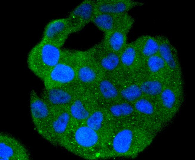
- Submitted by
- Invitrogen Antibodies (provider)
- Main image

- Experimental details
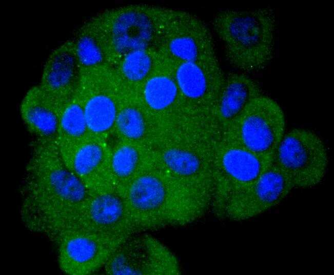
- Immunocytochemical analysis of S100A4 in MCF-7 cells using a S100A4 Monoclonal antibody (Product # MA5-32347) as seen in green. The nuclear counter stain is DAPI (blue). Cells were fixed in paraformaldehyde, permeabilised with 0.25% Triton X100/PBS.
- Submitted by
- Invitrogen Antibodies (provider)
- Main image

- Experimental details
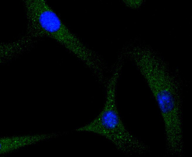
- Immunocytochemical analysis of S100A4 in NIH/3T3 cells using a S100A4 Monoclonal antibody (Product # MA5-32347) as seen in green. The nuclear counter stain is DAPI (blue). Cells were fixed in paraformaldehyde, permeabilised with 0.25% Triton X100/PBS.
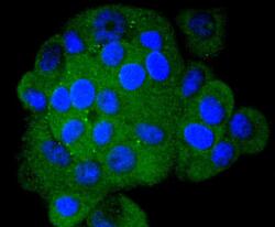
- Submitted by
- Invitrogen Antibodies (provider)
- Main image

- Experimental details
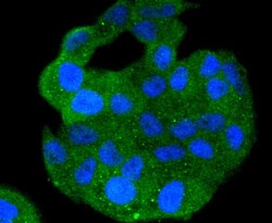
- Immunocytochemical analysis of S100A4 in Hela cells using a S100A4 Monoclonal antibody (Product # MA5-32347) as seen in green. The nuclear counter stain is DAPI (blue). Cells were fixed in paraformaldehyde, permeabilised with 0.25% Triton X100/PBS.

- Submitted by
- Invitrogen Antibodies (provider)
- Main image

- Experimental details
- Immunocytochemical analysis of S100A4 in NIH/3T3 cells using a S100A4 Monoclonal antibody (Product # MA5-32347) as seen in green. The nuclear counter stain is DAPI (blue). Cells were fixed in paraformaldehyde, permeabilised with 0.25% Triton X100/PBS.

- Submitted by
- Invitrogen Antibodies (provider)
- Main image
- Experimental details
- Immunocytochemical analysis of S100A4 in Hela cells using a S100A4 Monoclonal antibody (Product # MA5-32347) as seen in green. The nuclear counter stain is DAPI (blue). Cells were fixed in paraformaldehyde, permeabilised with 0.25% Triton X100/PBS.
- Submitted by
- Invitrogen Antibodies (provider)
- Main image
- Experimental details
- Immunocytochemical analysis of S100A4 in MCF-7 cells using a S100A4 Monoclonal antibody (Product # MA5-32347) as seen in green. The nuclear counter stain is DAPI (blue). Cells were fixed in paraformaldehyde, permeabilised with 0.25% Triton X100/PBS.

Supportive validation
- Submitted by
- Invitrogen Antibodies (provider)
- Main image
- Experimental details
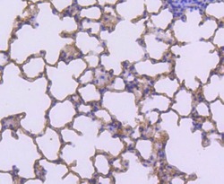
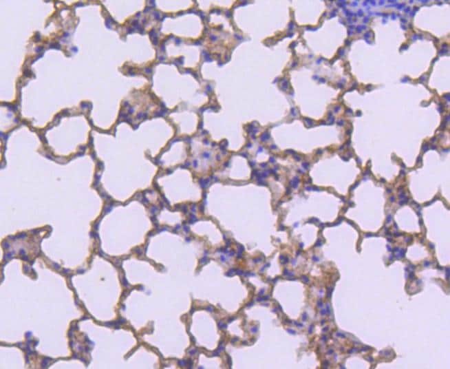
- Immunohistochemical analysis of S100A4 of paraffin-embedded Mouse lung tissue using a S100A4 Monoclonal antibody (Product #MA5-32347). Counter stained with hematoxylin.
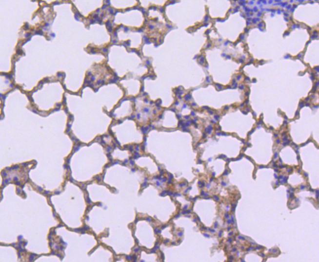
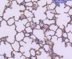
- Submitted by
- Invitrogen Antibodies (provider)
- Main image

- Experimental details
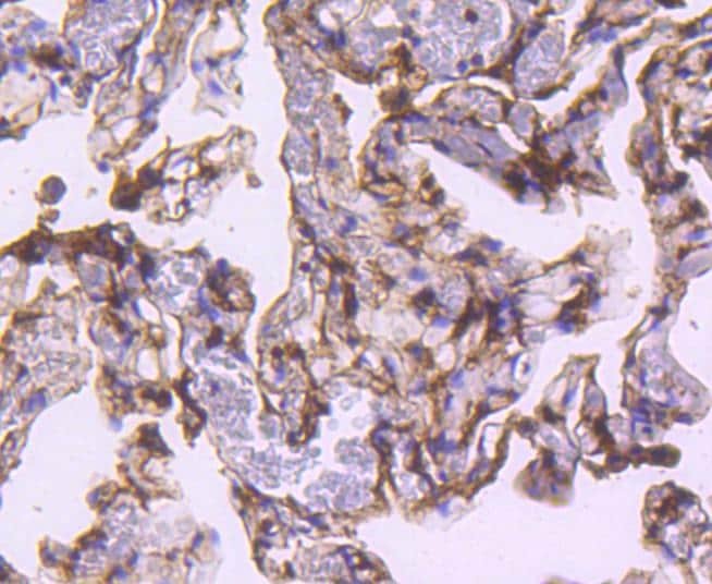
- Immunohistochemical analysis of S100A4 of paraffin-embedded Human lung tissue using a S100A4 Monoclonal antibody (Product #MA5-32347). Counter stained with hematoxylin.

- Submitted by
- Invitrogen Antibodies (provider)
- Main image
- Experimental details
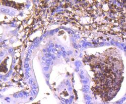
- Immunohistochemical analysis of S100A4 of paraffin-embedded Human gastric carcinoma tissue using a S100A4 Monoclonal antibody (Product #MA5-32347). Counter stained with hematoxylin.

- Submitted by
- Invitrogen Antibodies (provider)
- Main image
- Experimental details
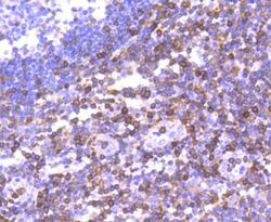
- Immunohistochemical analysis of S100A4 of paraffin-embedded Human tonsil tissue using a S100A4 Monoclonal antibody (Product #MA5-32347). Counter stained with hematoxylin.
- Submitted by
- Invitrogen Antibodies (provider)
- Main image
- Experimental details
- Immunohistochemical analysis of S100A4 of paraffin-embedded Mouse lung tissue using a S100A4 Monoclonal antibody (Product #MA5-32347). Counter stained with hematoxylin.
- Submitted by
- Invitrogen Antibodies (provider)
- Main image

- Experimental details
- Immunohistochemical analysis of S100A4 of paraffin-embedded Human lung tissue using a S100A4 Monoclonal antibody (Product #MA5-32347). Counter stained with hematoxylin.
- Submitted by
- Invitrogen Antibodies (provider)
- Main image

- Experimental details
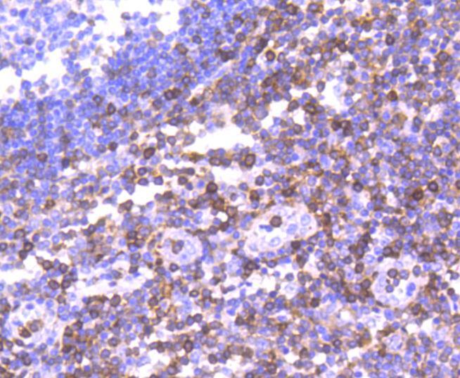
- Immunohistochemical analysis of S100A4 of paraffin-embedded Human tonsil tissue using a S100A4 Monoclonal antibody (Product #MA5-32347). Counter stained with hematoxylin.

Supportive validation
- Submitted by
- Invitrogen Antibodies (provider)
- Main image
- Experimental details
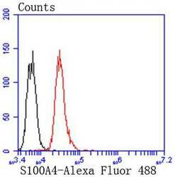
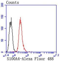
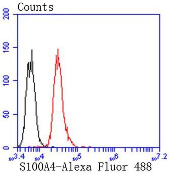
- Flow Cytometric analysis of S100A4 in Jurkat cells using a S100A4 Monoclonal Antibody (Product # MA5-32347) at a dilution of 1:50, as seen in red compared with an unlabelled control (cells without incubation with primary antibody; black). Alexa Fluor 488-conjugated goat anti rabbit IgG was used as the secondary antibody.

- Submitted by
- Invitrogen Antibodies (provider)
- Main image
- Experimental details
- Flow Cytometric analysis of S100A4 in Jurkat cells using a S100A4 Monoclonal Antibody (Product # MA5-32347) at a dilution of 1:50, as seen in red compared with an unlabelled control (cells without incubation with primary antibody; black). Alexa Fluor 488-conjugated goat anti rabbit IgG was used as the secondary antibody.
Supportive validation
- Submitted by
- Invitrogen Antibodies (provider)
- Main image
- Experimental details
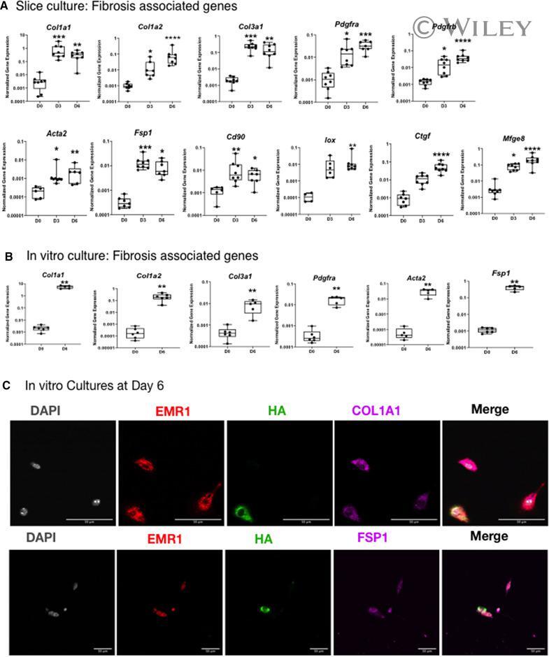
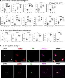
- 4 Figure Gene expression analysis of Kupffer cells cultured up to 6 days in slice cultures and in vitro cultures revealed upregulation of fibrosis-associated genes. (A and B) Emr1-Cre +/- ::RiboTag +/- mice livers were sliced and cultured (A) or processed to isolate non-parenchymal cells and cultured in vitro (B) for up to 6 days. (extended data in Supporting Information Figure 2). Gene expression was analyzed by qPCR at the indicated time points. (C) Representative immunofluorescence images indicating COL1A1 or FSP1 expression on HA labeled cells in in vitro cultures. Gray: Nuclei, Red: EMR1, Green: HA (RiboTag), Magenta: COL1A1, FSP1. All images 40x magnification Bar 50um. Data represent eight independent experiments with n = 1 per experiment in (A), six independent experiments with n = 1 per experiment in (B), and three independent experiments with n = 1 per experiment in (C). (A and B) In box and whisker plots, the middle line indicates the median, bars indicate the minimum to maximum. Dots: individual mice. *P