Explore
 Validate
 Learn
Learn Western blot
Antibody data
- Antibody Data
- Antigen structure
- References [0]
- Comments [0]
- Validations
- Western blot [3]
Submit
Validation data
Reference
Comment
Report error
- Product number
- AF4138 - Provider product page

- Provider
- R&D Systems
- Product name
- Human/Mouse S100A4 Antibody
- Antibody type
- Polyclonal
- Description
- Antigen Affinity-purified. Detects human and mouse S100A4 in direct ELISAs and Western blots. In direct ELISAs, less than 1% cross-reactivity with recombinant mouse (rm) S100A8, rmS100A6, rmS100A9, rmS100A10, rmS100A13, rmS100A16, recombinant human (rh) S100A1, rhS100A7, rhS100A11, rhS100B, and rhS100P is observed.
- Reactivity
- Human, Mouse
- Host
- Sheep
- Conjugate
- Unconjugated
- Antigen sequence
P07091- Isotype
- IgG
- Vial size
- 100 ug
- Concentration
- LYOPH
- Storage
- Use a manual defrost freezer and avoid repeated freeze-thaw cycles. 12 months from date of receipt, -20 to -70 °C as supplied. 1 month, 2 to 8 °C under sterile conditions after reconstitution. 6 months, -20 to -70 °C under sterile conditions after reconstitution.
No comments: Submit comment
Supportive validation
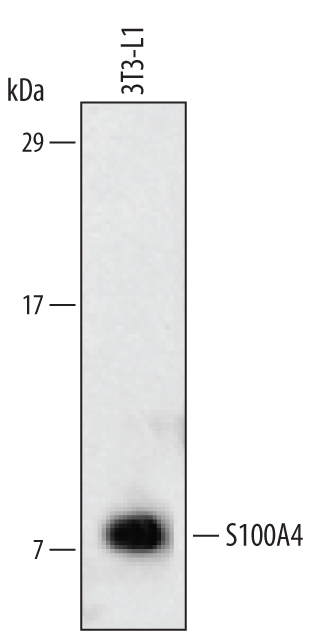
- Submitted by
- R&D Systems (provider)
- Main image

- Experimental details
- Detection of Mouse S100A4 by Simple WesternTM. Simple Western lane view shows lysates of 3T3-L1 mouse embryonic fibroblast adipose-like cell line, loaded at 0.2 mg/mL. A specific band was detected for S100A4 at approximately 12 kDa (as indicated) using 10 µg/mL of Sheep Anti-Human/Mouse S100A4 Antigen Affinity-purified Polyclonal Antibody (Catalog # AF4138) followed by 1:50 dilution of HRP-conjugated Anti-Sheep IgG Secondary Antibody (Catalog # HAF016). This experiment was conducted under reducing conditions and using the 12-230 kDa separation system.

- Submitted by
- R&D Systems (provider)
- Main image
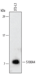
- Experimental details
- Detection of Mouse S100A4 by Western Blot. Western blot shows lysates of 3T3-L1 mouse embryonic fibroblast adipose-like cell line. PVDF membrane was probed with 1 µg/mL of Sheep Anti-Human/Mouse S100A4 Antigen Affinity-purified Polyclonal Antibody (Catalog # AF4138) followed by HRP-conjugated Anti-Sheep IgG Secondary Antibody (Catalog # HAF016). A specific band was detected for S100A4 at approximately 11 kDa (as indicated). This experiment was conducted under reducing conditions and using Immunoblot Buffer Group 2.
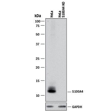
- Submitted by
- R&D Systems (provider)
- Main image

- Experimental details
- Western Blot Shows Sheep Anti-Human/Mouse S100A4 Antigen Affinity-purified Polyclonal Antibody Specificity by Using Knockout Cell Line. Western blot shows lysates of HeLa human cervical epithelial carcinoma parental cell line and S100A4 knockout HeLa cell line (KO). PVDF membrane was probed with 1 µg/mL of Sheep Anti-Human/Mouse S100A4 Antigen Affinity-purified Polyclonal Antibody (Catalog # AF4138) followed by HRP-conjugated Anti-Sheep IgG Secondary Antibody (Catalog # HAF016). A specific band was detected for S100A4 at approximately 12 kDa (as indicated) in the parental HeLa cell line, but is not detectable in knockout HeLa cell line. GAPDH (Catalog # AF5718) is shown as a loading control. This experiment was conducted under reducing conditions and using Immunoblot Buffer Group 1.