Explore
 Validate
 Learn
LearnPB9156
antibody from Boster Biological Technology
Targeting: PROM1
AC133, CD133, CORD12, MCDR2, PROML1, RP41, STGD4
 Western blot
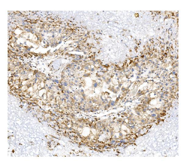
 Immunohistochemistry
Antibody data
- Antibody Data
- Antigen structure
- References [3]
- Comments [0]
- Validations
- Western blot [1]
Submit
Validation data
Reference
Comment
Report error
- Product number
- PB9156 - Provider product page

- Provider
- Boster Biological Technology
- Product name
- Anti-PROM1 Antibody Picoband™
- Antibody type
- Polyclonal
- Description
- Polyclonal antibody for CD133/PROM1 detection. Host: Rabbit.Size: 100μg/vial. Tested applications: WB, IHC-P, FCM. Reactive species: Human. CD133/PROM1 information: Molecular Weight: 97202 MW; Subcellular Localization: Apical cell membrane ; Multi- pass membrane protein . Cell projection, microvillus membrane ; Multi-pass membrane protein . Cell projection, cilium, photoreceptor outer segment . Endoplasmic reticulum. Endoplasmic reticulum-Golgi intermediate compartment. Found in extracellular membrane particles in various body fluids such as cerebrospinal fluid, saliva, seminal fluid and urine; Tissue Specificity: Isoform 1 is selectively expressed on CD34 hematopoietic stem and progenitor cells in adult and fetal bone marrow, fetal liver, cord blood and adult peripheral blood. Isoform 1 is not detected on other blood cells. Isoform 1 is also expressed in a number of non-lymphoid tissues including retina, pancreas, placenta, kidney, liver, lung, brain and heart. Found in saliva within small membrane particles. Isoform 2 is predominantly expressed in fetal liver, skeletal muscle, kidney, and heart as well as adult pancreas, kidney, liver, lung, and placenta. Isoform 2 is highly expressed in fetal liver, low in bone marrow, and barely detectable in peripheral blood. Isoform 2 is expressed on hematopoietic stem cells and in epidermal basal cells (at protein level). Expressed in adult retina by rod and cone photoreceptor cells (at protein level).
- Reactivity
- Human
- Host
- Rabbit
- Vial size
- 100μg/vial
- Concentration
- Add 0.2ml of distilled water will yield a concentration of 500ug/ml.
- Storage
- At -20°C for one year. After reconstitution, at 4°C for one month. It can also be aliquoted and stored frozen at -20°C for a longer time. Avoid repeated freezing and thawing.
- Handling
- Add 0.2ml of distilled water will yield a concentration of 500ug/ml.
Submitted references The CD133(+)CXCR4(+) Colorectal Tumor Cells Promote Colorectal Cancer Progression by PI3K/AKT Signaling.
Untangling the response of bone tumor cells and bone forming cells to matrix stiffness and adhesion ligand density by means of hydrogels.
Stemness marker ALDH1A1 promotes tumor angiogenesis via retinoic acid/HIF-1α/VEGF signalling in MCF-7 breast cancer cells.
Guan S, Yang R, Wu S, Xu K, Yang C
Journal of interferon & cytokine research : the official journal of the International Society for Interferon and Cytokine Research 2022 May;42(5):195-202
Journal of interferon & cytokine research : the official journal of the International Society for Interferon and Cytokine Research 2022 May;42(5):195-202
Untangling the response of bone tumor cells and bone forming cells to matrix stiffness and adhesion ligand density by means of hydrogels.
Jiang T, Zhao J, Yu S, Mao Z, Gao C, Zhu Y, Mao C, Zheng L
Biomaterials 2019 Jan;188:130-143
Biomaterials 2019 Jan;188:130-143
Stemness marker ALDH1A1 promotes tumor angiogenesis via retinoic acid/HIF-1α/VEGF signalling in MCF-7 breast cancer cells.
Ciccone V, Terzuoli E, Donnini S, Giachetti A, Morbidelli L, Ziche M
Journal of experimental & clinical cancer research : CR 2018 Dec 12;37(1):311
Journal of experimental & clinical cancer research : CR 2018 Dec 12;37(1):311
No comments: Submit comment
Supportive validation
- Submitted by
- Boster Biological Technology (provider)
- Main image
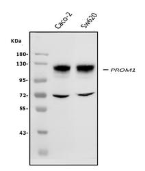
- Experimental details
- Western blot analysis of PROM1 using anti-PROM1 antibody (PB9156). Electrophoresis was performed on a 5-20% SDS-PAGE gel at 70V (Stacking gel) / 90V (Resolving gel) for 2-3 hours. The sample well of each lane was loaded with 50ug of sample under reducing conditions. Lane 1: human CACO-2 whole cell lysates, Lane 2: human SW620 whole cell lysates. After Electrophoresis, proteins were transferred to a Nitrocellulose membrane at 150mA for 50-90 minutes. Blocked the membrane with 5% Non-fat Milk/ TBS for 1.5 hour at RT. The membrane was incubated with rabbit anti-PROM1 antigen affinity purified polyclonal antibody (Catalog # PB9156) at 0.5 μg/mL overnight at 4°C, then washed with TBS-0.1%Tween 3 times with 5 minutes each and probed with a goat anti-rabbit IgG-HRP secondary antibody at a dilution of 1:10000 for 1.5 hour at RT. The signal is developed using an Enhanced Chemiluminescent detection (ECL) kit (Catalog # EK1002) with Tanon 5200 system. A specific band was detected for PROM1 at approximately 120KD. The expected band size for PROM1 is at 120KD.
- Additional image