Explore
 Validate
 Learn
Learn Western blot
 ELISA
ELISAAntibody data
- Antibody Data
- Antigen structure
- References [2]
- Comments [0]
- Validations
- Western blot [1]
Submit
Validation data
Reference
Comment
Report error
- Product number
- A07229-1 - Provider product page

- Provider
- Boster Biological Technology
- Product name
- Anti-OXCT1/SCOT Antibody Picoband™
- Antibody type
- Polyclonal
- Description
- Rabbit IgG polyclonal antibody for OXCT1/SCOT detection. Tested with WB, ICC/IF, FCM, Direct ELISA in Human;Mouse;Rat;Monkey.
- Reactivity
- Human, Mouse, Rat, Simian
- Host
- Rabbit
- Vial size
- 100μg/vial
- Concentration
- Add 0.2ml of distilled water will yield a concentration of 500ug/ml.
- Storage
- At -20°C for one year. After reconstitution, at 4°C for one month. It can also be aliquoted and stored frozen at -20°C for a longer time. Avoid repeated freezing and thawing.
- Handling
- Add 0.2ml of distilled water will yield a concentration of 500ug/ml.
Submitted references Tocopherol attenuates the oxidative stress of BMSCs by inhibiting ferroptosis through the PI3k/AKT/mTOR pathway.
Proanthocyanidin promotes functional recovery of spinal cord injury via inhibiting ferroptosis.
Lan D, Yao C, Li X, Liu H, Wang D, Wang Y, Qi S
Frontiers in bioengineering and biotechnology 2022;10:938520
Frontiers in bioengineering and biotechnology 2022;10:938520
Proanthocyanidin promotes functional recovery of spinal cord injury via inhibiting ferroptosis.
Zhou H, Yin C, Zhang Z, Tang H, Shen W, Zha X, Gao M, Sun J, Xu X, Chen Q
Journal of chemical neuroanatomy 2020 Sep;107:101807
Journal of chemical neuroanatomy 2020 Sep;107:101807
No comments: Submit comment
Supportive validation
- Submitted by
- Boster Biological Technology (provider)
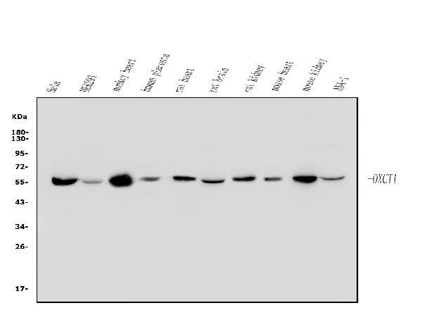
- Main image
- Experimental details
- Western blot analysis of OXCT1/SCOT using anti-OXCT1/SCOT antibody (A07229-1). Electrophoresis was performed on a 5-20% SDS-PAGE gel at 70V (Stacking gel) / 90V (Resolving gel) for 2-3 hours. The sample well of each lane was loaded with 50ug of sample under reducing conditions. Lane 1: human HELA whole cell lysates, Lane 2: human HEK293 whole cell lysates, Lane 3: monkey heart tissue lysates, Lane 4: human placenta tissue lysates, Lane 5: rat heart tissue lysates, Lane 6: rat brain tissue lysates, Lane 7: rat kidney tissue lysates, Lane 8: mouse heart tissue lysates, Lane 9: mouse kidney tissue lysates, Lane 10: mouse ANA-1 whole cell lysates. After Electrophoresis, proteins were transferred to a Nitrocellulose membrane at 150mA for 50-90 minutes. Blocked the membrane with 5% Non-fat Milk/ TBS for 1.5 hour at RT. The membrane was incubated with rabbit anti-OXCT1/SCOT antigen affinity purified polyclonal antibody (Catalog # A07229-1) at 0.25 μg/mL overnight at 4°C, then washed with TBS-0.1%Tween 3 times with 5 minutes each and probed with a goat anti-rabbit IgG-HRP secondary antibody at a dilution of 1:5000 for 1.5 hour at RT. The signal is developed using an Enhanced Chemiluminescent detection (ECL) kit (Catalog # EK1002) with Tanon 5200 system. A specific band was detected for OXCT1/SCOT at approximately 56KD. The expected band size for OXCT1/SCOT is at 56KD.
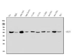
- Additional image