Explore
 Validate
 Learn
Learn Western blot
 Immunocytochemistry
Antibody data
- Antibody Data
- Antigen structure
- References [4]
- Comments [0]
- Validations
- Immunocytochemistry [4]
- Immunoprecipitation [1]
- Immunohistochemistry [2]
- Other assay [4]
Submit
Validation data
Reference
Comment
Report error
- Product number
- PA5-22168 - Provider product page

- Provider
- Invitrogen Antibodies
- Product name
- CaMKII delta Polyclonal Antibody
- Antibody type
- Polyclonal
- Antigen
- Recombinant full-length protein
- Description
- Recommended positive controls: 293T, A431, HeLa, HepG2, A549, H1299, HCT116, mouse brain, rat brain. Predicted reactivity: Mouse (100%), Rat (80%), Xenopus laevis (94%), Pig (100%), Rabbit (99%), Chicken (98%), Bovine (92%). Store product as a concentrated solution. Centrifuge briefly prior to opening the vial.
- Reactivity
- Human, Mouse, Rat, Rabbit
- Host
- Rabbit
- Isotype
- IgG
- Vial size
- 100 μL
- Concentration
- 0.66 mg/mL
- Storage
- Store at 4°C short term. For long term storage, store at -20°C, avoiding freeze/thaw cycles.
Submitted references Detrimental proarrhythmogenic interaction of Ca(2+)/calmodulin-dependent protein kinase II and Na(V)1.8 in heart failure.
Long-term effects of empagliflozin on excitation-contraction-coupling in human induced pluripotent stem cell cardiomyocytes.
Inhibition of calcium/calmodulin-dependent kinase II restores contraction and relaxation in isolated cardiac muscle from type 2 diabetic rats.
Beta-Adrenoceptor Stimulation Reveals Ca2+ Waves and Sarcoplasmic Reticulum Ca2+ Depletion in Left Ventricular Cardiomyocytes from Post-Infarction Rats with and without Heart Failure.
Bengel P, Dybkova N, Tirilomis P, Ahmad S, Hartmann N, A Mohamed B, Krekeler MC, Maurer W, Pabel S, Trum M, Mustroph J, Gummert J, Milting H, Wagner S, Ljubojevic-Holzer S, Toischer K, Maier LS, Hasenfuss G, Streckfuss-Bömeke K, Sossalla S
Nature communications 2021 Nov 15;12(1):6586
Nature communications 2021 Nov 15;12(1):6586
Long-term effects of empagliflozin on excitation-contraction-coupling in human induced pluripotent stem cell cardiomyocytes.
Pabel S, Reetz F, Dybkova N, Shomroni O, Salinas G, Mustroph J, Hammer KP, Hasenfuss G, Hamdani N, Maier LS, Streckfuss-Bömeke K, Sossalla S
Journal of molecular medicine (Berlin, Germany) 2020 Dec;98(12):1689-1700
Journal of molecular medicine (Berlin, Germany) 2020 Dec;98(12):1689-1700
Inhibition of calcium/calmodulin-dependent kinase II restores contraction and relaxation in isolated cardiac muscle from type 2 diabetic rats.
Daniels LJ, Wallace RS, Nicholson OM, Wilson GA, McDonald FJ, Jones PP, Baldi JC, Lamberts RR, Erickson JR
Cardiovascular diabetology 2018 Jun 14;17(1):89
Cardiovascular diabetology 2018 Jun 14;17(1):89
Beta-Adrenoceptor Stimulation Reveals Ca2+ Waves and Sarcoplasmic Reticulum Ca2+ Depletion in Left Ventricular Cardiomyocytes from Post-Infarction Rats with and without Heart Failure.
Sadredini M, Danielsen TK, Aronsen JM, Manotheepan R, Hougen K, Sjaastad I, Stokke MK
PloS one 2016;11(4):e0153887
PloS one 2016;11(4):e0153887
No comments: Submit comment
Supportive validation
- Submitted by
- Invitrogen Antibodies (provider)
- Main image

- Experimental details
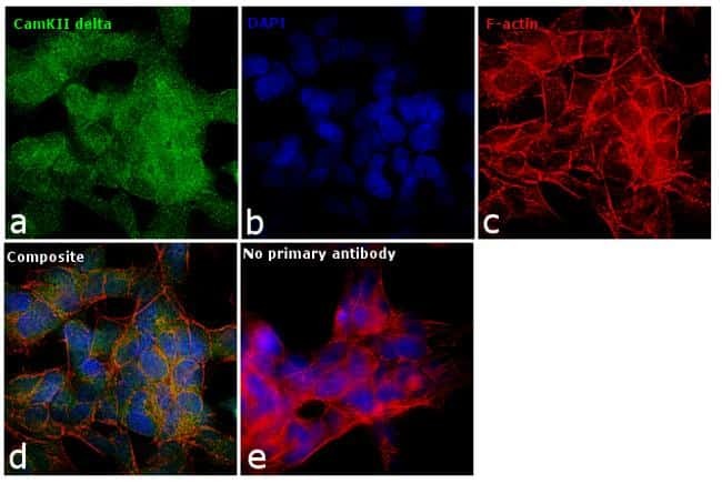
- Immunofluorescence analysis of CamKII delta was performed using 70% confluent log phase SH-SY5Y cells. The cells were fixed with 4% paraformaldehyde for 10 minutes, permeabilized with 0.1% Triton™ X-100 for 15 minutes, and blocked with 1% BSA for 1 hour at room temperature. The cells were labeled with CaMKII delta Polyclonal Antibody (Product # PA5-22168) at 5 µg/mL in 0.1% BSA, incubated at 4 degree Celsius overnight and then labeled with Goat anti-Rabbit IgG (H+L) Superclonal™ Secondary Antibody, Alexa Fluor® 488 conjugate (Product # A27034) at a dilution of 1:2000 for 45 minutes at room temperature (Panel a: green). Nuclei (Panel b: blue) were stained with SlowFade® Gold Antifade Mountant with DAPI (Product # S36938). F-actin (Panel c: red) was stained with Rhodamine Phalloidin (Product # R415, 1:300). Panel d represents the merged image showing predominant cytoplasmic localization. Panel e represents control cells with no primary antibody to assess background. The images were captured at 60X magnification.
- Submitted by
- Invitrogen Antibodies (provider)
- Main image

- Experimental details
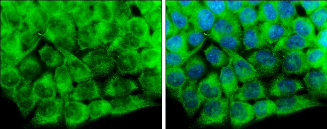
- Immunocytochemistry-Immunofluorescence analysis of CaMKII delta was performed in HCT116 cells fixed in 4% paraformaldehyde at RT for 15 min. Green: CaMKII delta Polyclonal Antibody (Product # PA5 22168) diluted at 1:500. Blue: Hoechst 33342 staining.

- Submitted by
- Invitrogen Antibodies (provider)
- Main image

- Experimental details
- Immunofluorescence analysis of CamKII delta was performed using 70% confluent log phase SH-SY5Y cells. The cells were fixed with 4% paraformaldehyde for 10 minutes, permeabilized with 0.1% Triton™ X-100 for 15 minutes, and blocked with 1% BSA for 1 hour at room temperature. The cells were labeled with CaMKII delta Polyclonal Antibody (Product # PA5-22168) at 5 µg/mL in 0.1% BSA, incubated at 4 degree Celsius overnight and then labeled with Goat anti-Rabbit IgG (Heavy Chain) Superclonal™ Secondary Antibody, Alexa Fluor® 488 conjugate (Product # A27034) at a dilution of 1:2000 for 45 minutes at room temperature (Panel a: green). Nuclei (Panel b: blue) were stained with SlowFade® Gold Antifade Mountant with DAPI (Product # S36938). F-actin (Panel c: red) was stained with Rhodamine Phalloidin (Product # R415, 1:300). Panel d represents the merged image showing predominant cytoplasmic localization. Panel e represents control cells with no primary antibody to assess background. The images were captured at 60X magnification.

- Submitted by
- Invitrogen Antibodies (provider)
- Main image

- Experimental details
- Immunocytochemistry-Immunofluorescence analysis of CaMKII delta was performed in HCT116 cells fixed in 4% paraformaldehyde at RT for 15 min. Green: CaMKII delta Polyclonal Antibody (Product # PA5 22168) diluted at 1:500. Blue: Hoechst 33342 staining.
Supportive validation
- Submitted by
- Invitrogen Antibodies (provider)
- Main image
- Experimental details
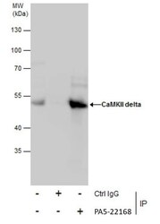
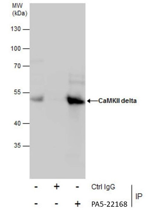
- Immunoprecipitation of CaMKII delta was performed in HepG2 whole cell extracts using 5 µg of CaMKII delta Polyclonal Antibody (Product # PA5-22168). Samples were transferred to a membrane and probed with CaMKII delta Polyclonal Antibody as a primary antibody and an HRP-conjugated anti-Rabbit IgG was used as a secondary antibody.

Supportive validation
- Submitted by
- Invitrogen Antibodies (provider)
- Main image
- Experimental details
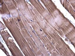
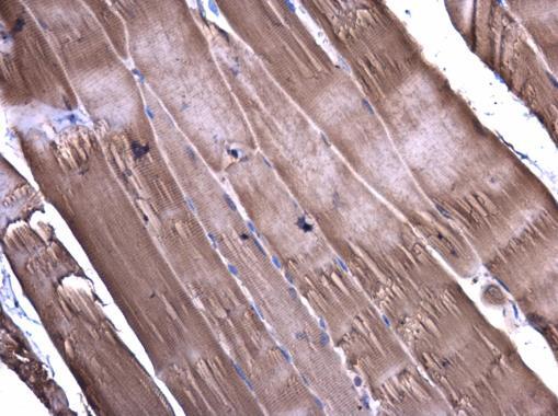
- Immunohistochemistry (Paraffin) analysis of CaMKII delta was performed in paraffin-embedded mouse muscle tissue using CaMKII delta Polyclonal Antibody (Product # PA5-22168) at a dilution of 1:400.
- Submitted by
- Invitrogen Antibodies (provider)
- Main image
- Experimental details
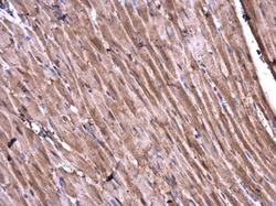
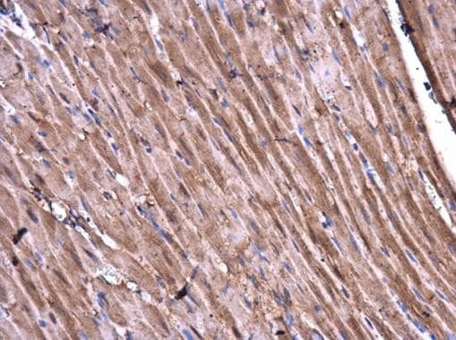
- Immunohistochemistry (Paraffin) analysis of CaMKII delta was performed in paraffin-embedded rat heart tissue using CaMKII delta Polyclonal Antibody (Product # PA5-22168) at a dilution of 1:400.
Supportive validation
- Submitted by
- Invitrogen Antibodies (provider)
- Main image
- Experimental details
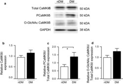
- Fig. 2 CaMKIIdelta activation is increased in right ventricular tissue from type 2 diabetic ZDF rats compared to non-diabetic controls. a Representative immunoblot of total CaMKIIdelta, Thr287 phosphorylated (PCaMKIIdelta) and GAPDH as a loading control in nDM and DM ZDF right ventricle tissue. b Quantification of total CaMKIIdelta levels in nDM (n = 8) and DM (n = 8) ZDF right ventricle tissue. c Quantification of PCaMKIIdelta levels in nDM (n = 8) and DM rats (n = 8). d Quantification of O-GlcNAc modified CaMKIIdelta levels in non-diabetic nDM and DM ZDF right ventricular tissue (DM) (n = 5). Data are mean +- SEM. *p < 0.05 for comparison of nDM and DM values (unpaired t-test)

- Submitted by
- Invitrogen Antibodies (provider)
- Main image

- Experimental details
- Immunoprecipitation of CaMKII delta was performed in HepG2 whole cell extracts using 5 µg of CaMKII delta Polyclonal Antibody (Product # PA5-22168). Samples were transferred to a membrane and probed with CaMKII delta Polyclonal Antibody as a primary antibody and an HRP-conjugated anti-Rabbit IgG was used as a secondary antibody.
- Submitted by
- Invitrogen Antibodies (provider)
- Main image
- Experimental details
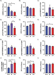
- Fig 6 Immunoblotting. Immunoblot analysis of key Ca 2+ handling proteins and phosphorylation was performed on tissue from left ventricles. SERCA2A abundance (A). PLB abundance (B) and phosphorylation on SER16 (C) and THR17 (D). NCX abundance (E). RyR abundance (F) with phosphorylation on SER2808 (G) and SER2814 (H). CaMKII abundance (I) and CaMKII phosphorylation on THR286 (J). PP1 (K) and PP2A (L) abundance. n heart = 6 for all analysis. *p

- Submitted by
- Invitrogen Antibodies (provider)
- Main image

- Experimental details
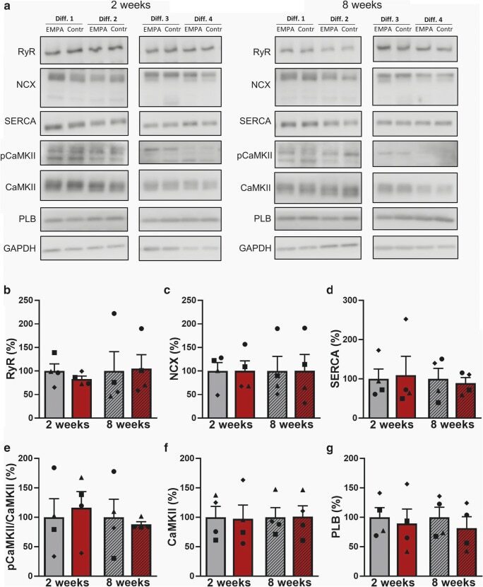
- Fig. 6 Western blot of EC-coupling proteins in human-induced pluripotent stem cell cardiomyocytes (iPSC-CM). ( a ) Representative original Western blots after treatment with empagliflozin (EMPA) or vehicle control (control) for 2 or 8 weeks from 4 differentiation experiments (Diff.) from 2 healthy donors. GAPDH was used as loading control. ( b ) Mean protein expression levels (normalized to the respective control group at 2 or 8 weeks) in iPSC-CM ( n = 4 differentiations, matched groups are displayed with matched individual symbols) and effects of 2 and 8 weeks treatment with empagliflozin (EMPA) on ryanodine-receptor type 2 (RyR2), ( c ) sodium-calcium exchanger (NCX), ( d ) sarcoplasmic reticulum Ca 2+ ATPase (SERCA), ( e ) phosphorylated Ca 2+ -/calmodulin-dependent protein kinase II (pCaMKII), ( f ) Ca 2+ -/calmodulin-dependent protein kinase II (CaMKII), and ( g ) phospholamban (PLB). Groups were statistically analyzed using two-way ANOVA with Sidak's test for multiple comparisons