 Explore
 Validate
 Learn
Learn Western blot
 Immunocytochemistry
Antibody data
- Antibody Data
- Antigen structure
- References [3]
- Comments [0]
- Validations
- Western blot [1]
- Immunohistochemistry [2]
Submit
Validation data
Reference
Comment
Report error
- Product number
- NB100-527 - Provider product page

- Provider
- Novus Biologicals
- Proper citation
- Novus Cat#NB100-527, RRID:AB_10003274
- Product name
- Rabbit Polyclonal VEGFR1/Flt-1 Antibody
- Antibody type
- Polyclonal
- Description
- Immunogen affinity purified. This antibody targets VEGFR-1 but has significant cross-reactivity with the VEGFR-2 protein.
- Reactivity
- Human
- Host
- Rabbit
- Isotype
- IgG
- Vial size
- 0.1 ml
- Concentration
- 1 mg/ml
- Storage
- Store at 4C. Do not freeze.
Submitted references Tanshinone IIA can inhibit MiaPaCa‑2 human pancreatic cancer cells by dual blockade of the Ras/Raf/MEK/ERK and PI3K/AKT/mTOR pathways.
Positive feedback between vascular endothelial growth factor-A and autotaxin in ovarian cancer cells.
Expression of periostin in human breast cancer.
Su CC
Oncology reports 2018 Nov;40(5):3102-3111
Oncology reports 2018 Nov;40(5):3102-3111
Positive feedback between vascular endothelial growth factor-A and autotaxin in ovarian cancer cells.
Ptaszynska MM, Pendrak ML, Bandle RW, Stracke ML, Roberts DD
Molecular cancer research : MCR 2008 Mar;6(3):352-63
Molecular cancer research : MCR 2008 Mar;6(3):352-63
Expression of periostin in human breast cancer.
Puglisi F, Puppin C, Pegolo E, Andreetta C, Pascoletti G, D'Aurizio F, Pandolfi M, Fasola G, Piga A, Damante G, Di Loreto C
Journal of clinical pathology 2008 Apr;61(4):494-8
Journal of clinical pathology 2008 Apr;61(4):494-8
No comments: Submit comment
Supportive validation
- Submitted by
- Novus Biologicals (provider)
- Main image

- Experimental details
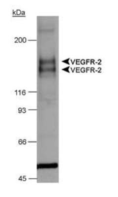
- Western Blot: VEGF R1/Flt-1 Antibody [NB100-527] - Chimeric CSF-1R/VEGFR-2 detection in transfected lysates.
Supportive validation
- Submitted by
- Novus Biologicals (provider)
- Main image
- Experimental details
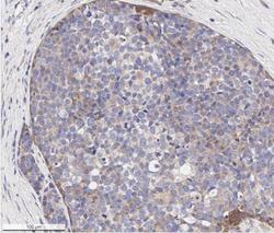
- Immunohistochemistry-Paraffin: VEGFR1/Flt-1 Antibody [NB100-527] - Analysis of FFPE human breast carcinoma tissue section using 1:500 dilution of VEGFR1/Flt-1 antibody on a Bond Rx autostainer (Leica Biosystems). The assay involved 20 minutes of heat induced antigen retrieval (HIER) with 10mM sodium citrate buffer (pH 6.0) and endogenous peroxidase quenching using peroxide block. The sections were incubated with primary antibody for 30 minutes. Bond Polymer Refine Detection (Leica Biosystems) and DAB were used for signal detection which followed counterstaining with hematoxylin. Whole slide scanning and capturing of representative images (20X) were performed using Aperio AT2 (Leica Biosystems). This VEGFR1/Flt-1 antibody generated an expected membrane cytoplasmic staining of VEGFR1 protein in the cancer cells (punctate appearance typical of receptors). The tumor stroma/stromal cells did not show VEGFR1/Flt-1immunopositivity.

- Submitted by
- Novus Biologicals (provider)
- Main image
- Experimental details
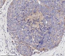
- Immunohistochemistry-Paraffin: VEGFR1/Flt-1 Antibody [NB100-527] - Analysis of a FFPE human breast carcinoma tissue section using 1:500 dilution of VEGFR1/Flt-1 antibody on a Bond Rx autostainer (Leica Biosystems). The assay involved 20 minutes of heat induced antigen retrieval (HIER) with 10mM sodium citrate buffer (pH 6.0) and endogenous peroxidase quenching using peroxide block. The sections were incubated with primary antibody for 30 minutes. Bond Polymer Refine Detection (Leica Biosystems) and DAB were used for signal detection which followed counterstaining with hematoxylin. Whole slide scanning and capturing of representative images (20X) were performed using Aperio AT2 (Leica Biosystems). This VEGFR1/Flt-1 antibody generated an expected membrane cytoplasmic staining of VEGFR1 protein in the cancer cells. The tumor stroma/stromal cells did not show VEGFR1/Flt-1immunopositivity. Staining was performed by Histowiz.
