Explore
 Validate
 Learn
Learn Western blot
 Immunohistochemistry
Antibody data
- Antibody Data
- Antigen structure
- References [0]
- Comments [0]
- Validations
- Immunohistochemistry [1]
Submit
Validation data
Reference
Comment
Report error
- Product number
- AMAb90704 - Provider product page

- Provider
- Atlas Antibodies
- Proper citation
- Atlas Antibodies Cat#AMAb90704, RRID:AB_2665637
- Product name
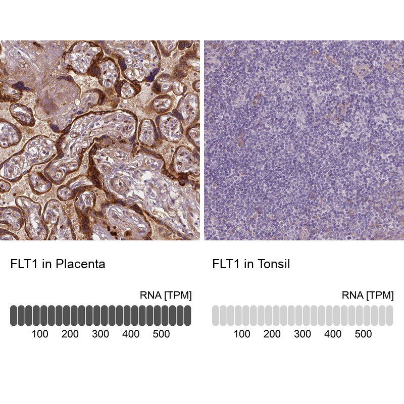
- Anti-FLT1
- Antibody type
- Monoclonal
- Description
- Monoclonal Antibody against Human FLT1, Clone ID: CL0345, Gene description: fms-related tyrosine kinase 1 (vascular endothelial growth factor/vascular permeability factor receptor), Alternative Gene Names: FLT, VEGFR1, Validated applications: IHC, WB, Uniprot ID: P17948, Storage: Store at +4°C for short term storage. Long time storage is recommended at -20°C.
- Reactivity
- Human
- Host
- Mouse
- Conjugate
- Unconjugated
- Isotype
- IgG
- Antibody clone number
- CL0345
- Vial size
- 100 µl
- Concentration
- 1.0 mg/ml
- Storage
- Store at +4°C for short term storage. Long time storage is recommended at -20°C.
- Handling
- The antibody solution should be gently mixed before use.
No comments: Submit comment
Supportive validation
- Submitted by
- Atlas Antibodies (provider)
- Enhanced method
- Orthogonal validation
- Main image
- Experimental details
- Immunohistochemistry analysis in human placenta and tonsil tissues using AMAb90704 antibody. Corresponding FLT1 RNA-seq data are presented for the same tissues.
- Sample type
- Human
- Protocol
- Protocol