Explore
 Validate
 Learn
Learn Western blot
Antibody data
- Antibody Data
- Antigen structure
- References [0]
- Comments [0]
- Validations
- Western blot [2]
- Immunohistochemistry [13]
Submit
Validation data
Reference
Comment
Report error
- Product number
- NBP1-91241 - Provider product page

- Provider
- Novus Biologicals
- Proper citation
- Novus Cat#NBP1-91241, RRID:AB_11034525
- Product name
- Rabbit Polyclonal SAMD9L Antibody
- Antibody type
- Polyclonal
- Description
- Immunogen affinity purified. Specificity of human SAMD9L antibody verified on a Protein Array containing target protein plus 383 other non-specific proteins.
- Reactivity
- Human
- Host
- Rabbit
- Isotype
- IgG
- Vial size
- 0.1 ml
- Storage
- Store at 4C short term. Aliquot and store at -20C long term. Avoid freeze-thaw cycles.
No comments: Submit comment
Supportive validation
- Submitted by
- Novus Biologicals (provider)
- Main image

- Experimental details
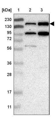
- Western Blot: SAMD9L Antibody [NBP1-91241] - Lane 1: Marker [kDa] 230, 130, 95, 72, 56, 36, 28, 17, 11 Lane 2: Human cell line RT-4 Lane 3: Human cell line U-251MG sp
- Submitted by
- Novus Biologicals (provider)
- Main image
- Experimental details
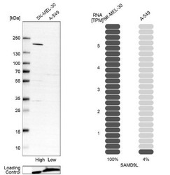
- Western Blot: SAMD9L Antibody [NBP1-91241] - Analysis in human cell line SK-MEL-30 and human cell line A-549.

Supportive validation
- Submitted by
- Novus Biologicals (provider)
- Main image
- Experimental details
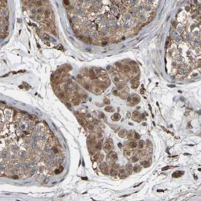
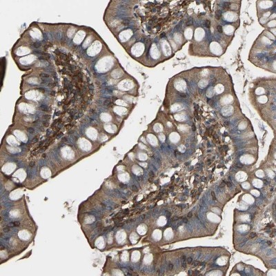
- Immunohistochemistry-Paraffin: SAMD9L Antibody [NBP1-91241] - Staining of human small intestine shows moderate cytoplasmic and membranous positivity in glandular cells.
- Submitted by
- Novus Biologicals (provider)
- Main image
- Experimental details
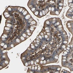
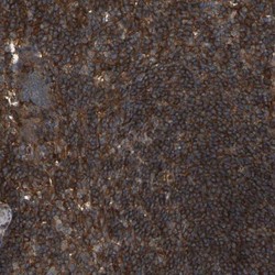
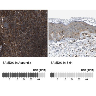
- Immunohistochemistry-Paraffin: SAMD9L Antibody [NBP1-91241] - Staining of human appendix shows high expression.

- Submitted by
- Novus Biologicals (provider)
- Main image
- Experimental details
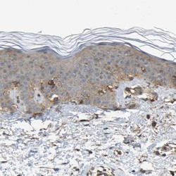
- Immunohistochemistry-Paraffin: SAMD9L Antibody [NBP1-91241] - Staining of human skin shows low expression as expected.

- Submitted by
- Novus Biologicals (provider)
- Main image
- Experimental details
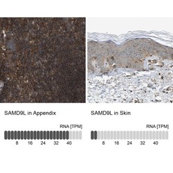
- Immunohistochemistry-Paraffin: SAMD9L Antibody [NBP1-91241] - Staining in human appendix and skin tissues using anti-SAMD9L antibody. Corresponding SAMD9L RNA-seq data are presented for the same tissues.
- Submitted by
- Novus Biologicals (provider)
- Main image
- Experimental details
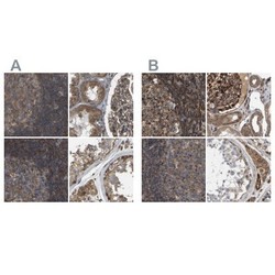
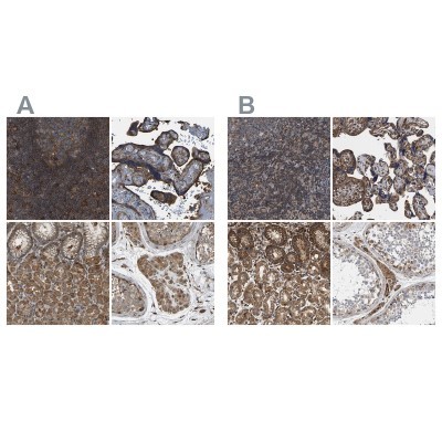
- Immunohistochemistry-Paraffin: SAMD9L Antibody [NBP1-91241] - Staining of human appendix, kidney, lymph node and testis using Anti-SAMD9L antibody NBP1-91241 (A) shows similar protein distribution across tissues to independent antibody NBP1-91242 (B).
- Submitted by
- Novus Biologicals (provider)
- Main image
- Experimental details
- Immunohistochemistry-Paraffin: SAMD9L Antibody [NBP1-91241] - Staining of human lymph node.
- Submitted by
- Novus Biologicals (provider)
- Main image

- Experimental details
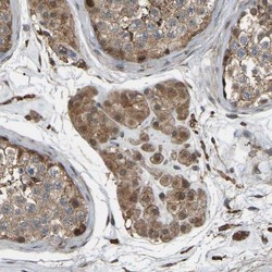
- Immunohistochemistry-Paraffin: SAMD9L Antibody [NBP1-91241] - Staining of human testis.
- Submitted by
- Novus Biologicals (provider)
- Main image
- Experimental details
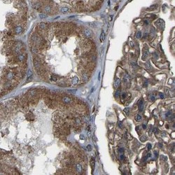
- Immunohistochemistry-Paraffin: SAMD9L Antibody [NBP1-91241] - Staining of human kidney.

- Submitted by
- Novus Biologicals (provider)
- Main image

- Experimental details
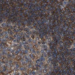
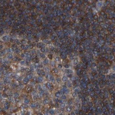
- Immunohistochemistry-Paraffin: SAMD9L Antibody [NBP1-91241] - Staining of human lymph node shows strong cytoplasmic positivity in non-germinal center cells.

- Submitted by
- Novus Biologicals (provider)
- Main image

- Experimental details
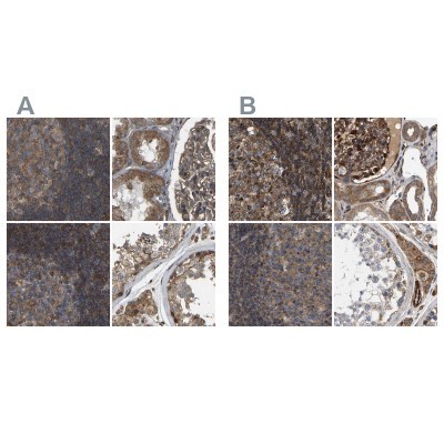
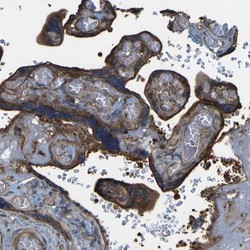
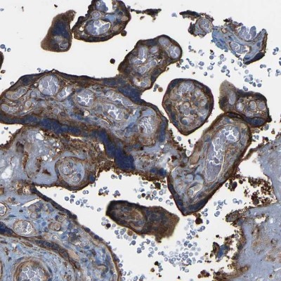
- Immunohistochemistry-Paraffin: SAMD9L Antibody [NBP1-91241] - Staining of human lymph node, placenta, stomach and testis using Anti-SAMD9L antibody NBP1-91241 (A) shows similar protein distribution across tissues to independent antibody NBP1-91242 (B).
- Submitted by
- Novus Biologicals (provider)
- Main image
- Experimental details
- Immunohistochemistry-Paraffin: SAMD9L Antibody [NBP1-91241] - Staining of human placenta shows strong granular cytoplasmic positivity in trophoblastic cells.
- Submitted by
- Novus Biologicals (provider)
- Main image
- Experimental details
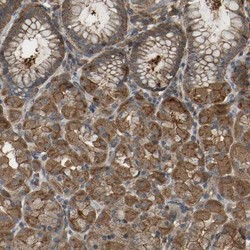
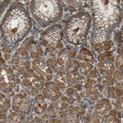
- Immunohistochemistry-Paraffin: SAMD9L Antibody [NBP1-91241] - Staining of human stomach shows strong cytoplasmic positivity in glandular cells.
- Submitted by
- Novus Biologicals (provider)
- Main image
- Experimental details
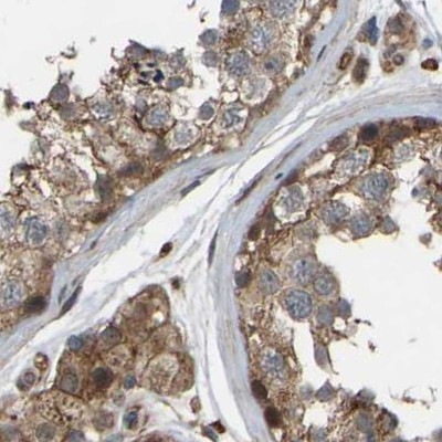
- Immunohistochemistry-Paraffin: SAMD9L Antibody [NBP1-91241] - Staining of human testis shows moderate cytoplasmic positivity in Leydig cells.