 Explore
 Validate
 Learn
Learn Western blot
 Immunocytochemistry
Antibody data
- Antibody Data
- Antigen structure
- References [4]
- Comments [0]
- Validations
- Immunocytochemistry [1]
- Immunohistochemistry [1]
- Other assay [3]
Submit
Validation data
Reference
Comment
Report error
- Product number
- PA5-30073 - Provider product page

- Provider
- Invitrogen Antibodies
- Product name
- ISG20 Polyclonal Antibody
- Antibody type
- Polyclonal
- Antigen
- Recombinant full-length protein
- Description
- Recommended positive controls: Raji, mouse spleen. Predicted reactivity: Mouse (83%), Rat (82%), Pig (81%), Rhesus Monkey (97%). Store product as a concentrated solution. Centrifuge briefly prior to opening the vial.
- Reactivity
- Human, Mouse
- Host
- Rabbit
- Isotype
- IgG
- Vial size
- 100 μL
- Concentration
- 0.44 mg/mL
- Storage
- Store at 4°C short term. For long term storage, store at -20°C, avoiding freeze/thaw cycles.
Submitted references USP22 controls type III interferon signaling and SARS-CoV-2 infection through activation of STING.
ISG15-dependent activation of the sensor MDA5 is antagonized by the SARS-CoV-2 papain-like protease to evade host innate immunity.
ISG15-dependent Activation of the RNA Sensor MDA5 and its Antagonism by the SARS-CoV-2 papain-like protease.
Interferon-Stimulated Gene (ISG)-Expression Screening Reveals the Specific Antibunyaviral Activity of ISG20.
Karlowitz R, Stanifer ML, Roedig J, Andrieux G, Bojkova D, Bechtel M, Smith S, Kowald L, Schubert R, Boerries M, Cinatl J Jr, Boulant S, van Wijk SJL
Cell death & disease 2022 Aug 6;13(8):684
Cell death & disease 2022 Aug 6;13(8):684
ISG15-dependent activation of the sensor MDA5 is antagonized by the SARS-CoV-2 papain-like protease to evade host innate immunity.
Liu G, Lee JH, Parker ZM, Acharya D, Chiang JJ, van Gent M, Riedl W, Davis-Gardner ME, Wies E, Chiang C, Gack MU
Nature microbiology 2021 Apr;6(4):467-478
Nature microbiology 2021 Apr;6(4):467-478
ISG15-dependent Activation of the RNA Sensor MDA5 and its Antagonism by the SARS-CoV-2 papain-like protease.
Liu G, Lee JH, Parker ZM, Acharya D, Chiang JJ, van Gent M, Riedl W, Davis-Gardner ME, Wies E, Chiang C, Gack MU
bioRxiv : the preprint server for biology 2020 Oct 27;
bioRxiv : the preprint server for biology 2020 Oct 27;
Interferon-Stimulated Gene (ISG)-Expression Screening Reveals the Specific Antibunyaviral Activity of ISG20.
Feng J, Wickenhagen A, Turnbull ML, Rezelj VV, Kreher F, Tilston-Lunel NL, Slack GS, Brennan B, Koudriakova E, Shaw AE, Rihn SJ, Rice CM, Bieniasz PD, Elliott RM, Shi X, Wilson SJ
Journal of virology 2018 Jul 1;92(13)
Journal of virology 2018 Jul 1;92(13)
No comments: Submit comment
Supportive validation
- Submitted by
- Invitrogen Antibodies (provider)
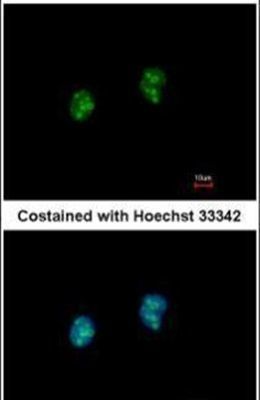
- Main image

- Experimental details
- Immunofluorescent analysis of ISG20 in paraformaldehyde-fixed HeLa cells using an ISG20 polyclonal antibody (Product # PA5-30073) at a 1:500 dilution.
Supportive validation
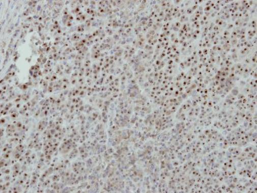
- Submitted by
- Invitrogen Antibodies (provider)
- Main image
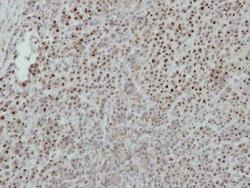
- Experimental details
- Immunohistochemical analysis of paraffin-embedded BT483 xenograft, using ISG20 (Product # PA5-30073) antibody at 1:500 dilution. Antigen Retrieval: Citrate buffer, pH 6.0, 15 min.
Supportive validation
- Submitted by
- Invitrogen Antibodies (provider)
- Main image
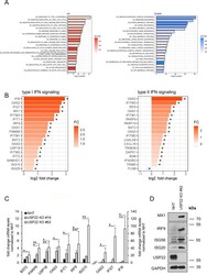
- Experimental details
- Loss of USP22 specifically enriches for genes involved in IFN signaling and response to viral infection. A Bar plot showing the top-20 regulated GO terms in two independent single-cell HT-29 USP22 CRISPR/Cas9 KO clones (#16 and #62) compared to control (NHT) HT-29 cells. Color code represents the number of annotated genes within each gene set. B Heatmap of differentially expressed genes contributing to the GO terms response to type I IFN (left) and type II signaling (right). Color code represents the log2 fold change compared to NHT. Note that due to lack of annotation and overlapping ISGs between type I/II and type III IFNs, response to type III IFN as GO term was not included. C Basal mRNA expression levels of GO- enriched genes related to IFN signaling in control (NHT) and two independent USP22 KO HT-29 single clones using qRT-PCR. Gene expression was normalized against 28S mRNA and is presented as x-fold mRNA expression compared to NHT. Mean and SD of three independent experiments in triplicate are shown. * P < 0.05; ** P < 0.01, *** P < 0.001, n.s. not significant. D Western blot analysis of basal MX1, IRF9, ISG56, ISG20, and USP22 expression levels in control and USP22 KO HT-29 cells (clone USP22 KO #62). GAPDH served as loading control. Representative blots of at least two different independent experiments are shown.

- Submitted by
- Invitrogen Antibodies (provider)
- Main image
- Experimental details
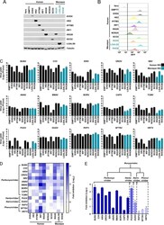
- FIG 3 Antibunyaviral activities of candidate ISGs against a panel of divergent bunyaviruses. (A) ISG expression in 10 Vero-ISG-expressing cell lines in addition to ""empty vector"" and naive Vero cell controls was assessed using Western blotting (assays were not established for NOS2A and RHOB). (B) For the ISGs in panel A, lentiviral vector-encoded TagRFP expression was monitored by flow cytometry. (C) Infectious virus yields from 10 Vero-ISG-expressing cell lines. Cells were infected with 15 wild-type bunyaviruses representing four families. Supernatants were collected, and virus yields were determined on Vero or BHK-21 cells. The MOIs and time points of supernatant harvest were as follows: for BUNV, CVV, KRIV, SBV, DUGV, RVFV, and SFTSV, MOI of 0.001 and 72 hpi; for OROV, MOI of 0.001 and 48 hpi; for ANAV, BMAV, BORV, CAPV, and TCMV, MOI of 0.01 and 48 hpi; for PUUV, MOI of 0.1 and 48 hpi; and for HRTV, MOI of 0.01 and 72 hpi. EMPTY, Vero cells transduced with empty SCRPSY lentiviral stocks. (D) Heat map analysis (generated by GraphPad Prism 7) of the effects of 10 ISGs on the yields of 15 bunyaviruses. (E) Summary of human ISG20's antiviral effects on 15 bunyaviruses.
- Submitted by
- Invitrogen Antibodies (provider)
- Main image
- Experimental details
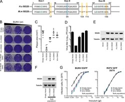
- FIG 4 A single point mutation in the active site (ISG20 D94G ) abolishes the antibunyaviral activity of ISG20. (A) Schematic representation of the three exonuclease active motifs (Exo I, II, and III) of ISG20 (shown in gray boxes). The conserved amino acid residues (from the DEDDh family) are highlighted in green. The Asp94 residue (shown in green) of wild-type ISG20 was replaced with Gly to generate the catalytically inactive mutant ISG20 D94G . The four amino acid differences between the human (H.s) and macaque (M.m) ISG20 proteins are highlighted with yellow boxes and red letters. (B) Plaque phenotypes of wt BUNV on Vero-EMPTY and Vero-ISG20/ISG20 D94G cells. (C and D) Quantification of plaque areas (C) and virus titers (D) of BUNV on Vero-EMPTY and Vero-ISG20/ISG20 D94G cells. (E) Western blot showing ISG20/ISG20 D94G protein expression. Cell lysates were probed with an anti-ISG20 polyclonal antibody. Tubulin served as a sample loading control. (F) Same as panel E, but using bulk populations of Vero cells expressing human and macaque ISG20. (G) Titration curves for the bulk populations from panel F infected with BUNV-EGFP ( 46 ) or single-cycle RVFV ( 57 ). Means and standard deviations (SD) for triplicate titrations are shown.
