Explore
 Validate
 Learn
Learn Western blot
Antibody data
- Antibody Data
- Antigen structure
- References [0]
- Comments [0]
- Validations
- Western blot [2]
- Immunocytochemistry [2]
- Immunohistochemistry [2]
- Flow cytometry [1]
Submit
Validation data
Reference
Comment
Report error
- Product number
- AGR-021-25UL - Provider product page

- Provider
- Invitrogen Antibodies
- Product name
- GLP1R (extracellular) Polyclonal Antibody
- Antibody type
- Polyclonal
- Antigen
- Other
- Reactivity
- Human, Mouse, Rat
- Host
- Rabbit
- Isotype
- IgG
- Vial size
- 25 µL
- Concentration
- 0.8 mg/mL
- Storage
- -20° C, Avoid Freeze/Thaw Cycles
No comments: Submit comment
Supportive validation
- Submitted by
- Invitrogen Antibodies (provider)
- Main image
- Experimental details
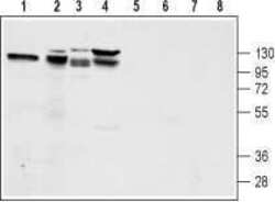
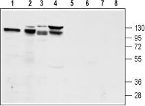
- Western blot analysis of rat pancreas lysate (lanes 1 and 5), mouse preadipocyte 3T3-L1 lysate (lanes 2 and 6), rat pancreatic islet cell line RIN-5F lysate (lanes 3 and 7) and human pancreatic carcinoma PANC-1 lysate (lanes 4 and 8): - 1-4. Anti-GLP1R (extracellular) Antibody (#AGR-021), (1:200).5-8. Anti-GLP1R (extracellular) Antibody , preincubated with GLP1R (extracellular) Blocking Peptide (#BLP-GR021).
- Submitted by
- Invitrogen Antibodies (provider)
- Main image
- Experimental details
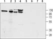
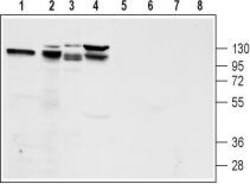
- Western blot analysis of rat pancreas lysate (lanes 1 and 5), mouse preadipocyte 3T3-L1 lysate (lanes 2 and 6), rat pancreatic islet cell line RIN-5F lysate (lanes 3 and 7) and human pancreatic carcinoma PANC-1 lysate (lanes 4 and 8): - 1-4. Anti-GLP1R (extracellular) Antibody (#AGR-021), (1:200).5-8. Anti-GLP1R (extracellular) Antibody , preincubated with GLP1R (extracellular) Blocking Peptide (#BLP-GR021).
Supportive validation
- Submitted by
- Invitrogen Antibodies (provider)
- Main image

- Experimental details
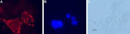
- Expression of Glucagon-like peptide 1 receptor in rat pancreatic islet cells - Cell surface detection of Glucagon-like peptide 1 receptor in live intact rat RIN-5F pancreatic islet cells. A. Cells were stained with Anti-GLP1R (extracellular) Antibody (#AGR-021), (1:100), followed by goat Anti-rabbit-AlexaFluor-594 secondary Antibody (red). B. Cell nuclei were visualized with the membrane-permeable DNA dye Hoechst 33342 (blue staining). C. Live view of the cells.
- Submitted by
- Invitrogen Antibodies (provider)
- Main image
- Experimental details
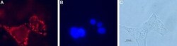
- Expression of Glucagon-like peptide 1 receptor in rat pancreatic islet cells - Cell surface detection of Glucagon-like peptide 1 receptor in live intact rat RIN-5F pancreatic islet cells. A. Cells were stained with Anti-GLP1R (extracellular) Antibody (#AGR-021), (1:100), followed by goat Anti-rabbit-AlexaFluor-594 secondary Antibody (red). B. Cell nuclei were visualized with the membrane-permeable DNA dye Hoechst 33342 (blue staining). C. Live view of the cells.

Supportive validation
- Submitted by
- Invitrogen Antibodies (provider)
- Main image

- Experimental details
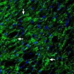
- Expression of Glucagon-like peptide 1 receptor in rat hypothalamus - Immunohistochemical staining of rat hypothalamus with Anti-GLP1R (extracellular) Antibody (#AGR-021), (1:200). Glucagon-like peptide 1 receptor (green) appears in the lateral hypothalamic region axonal profiles (vertical arrow) and nerve cell profiles (horizontal arrows). DAPI is used as the counterstain (blue).
- Submitted by
- Invitrogen Antibodies (provider)
- Main image

- Experimental details
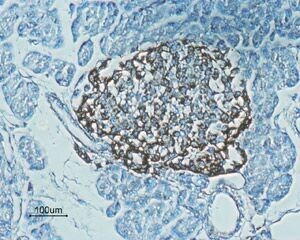
- Expression of Glucagon-like peptide 1 receptor in rat pancreas - Immunohistochemical staining of rat pancreas paraffin embedded sections using Anti-GLP1R (extracellular) Antibody (#AGR-021), (1:100). Staining (brown color) is present in endocrine cells of the Isles of Langerhans. Hematoxilin is used as the counterstain.
Supportive validation
- Submitted by
- Invitrogen Antibodies (provider)
- Main image
- Experimental details
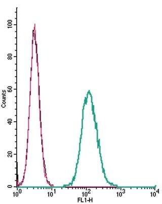
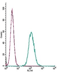
- Cell surface detectionofGlucagon-like peptide 1 receptorbyindirect flow cytometry in live intactmouse J774 macrophage cells: - (black line) cells. (red) Cells+ goat- Anti-rabbit-FITC. (green) Cells + Anti-GLP1R (extracellular) Antibody (#AGR-021), (2.5μg) + goat- Anti-rabbit-FITC.