Explore
 Validate
 Learn
Learn Western blot
 Other assay
Antibody data
- Antibody Data
- Antigen structure
- References [1]
- Comments [0]
- Validations
- Other assay [2]
Submit
Validation data
Reference
Comment
Report error
- Product number
- PA1-84457 - Provider product page

- Provider
- Invitrogen Antibodies
- Product name
- Calcitonin Receptor Polyclonal Antibody
- Antibody type
- Polyclonal
- Antigen
- Synthetic peptide
- Description
- When using two or more Super Bright dye-conjugated antibodies in a staining panel, it is recommended to use Super Bright Complete Staining Buffer (Product # SB-4401) to minimize any non-specific polymer interactions. Please refer to the datasheet for Super Bright Staining Buffer for more information.
- Reactivity
- Human, Mouse, Rat
- Host
- Rabbit
- Isotype
- IgG
- Vial size
- 100 µg
- Concentration
- 1 mg/mL
- Storage
- 4°C or -20°C if preferred
Submitted references Role of microglial amylin receptors in mediating beta amyloid (Aβ)-induced inflammation.
Fu W, Vukojevic V, Patel A, Soudy R, MacTavish D, Westaway D, Kaur K, Goncharuk V, Jhamandas J
Journal of neuroinflammation 2017 Oct 6;14(1):199
Journal of neuroinflammation 2017 Oct 6;14(1):199
No comments: Submit comment
Supportive validation
- Submitted by
- Invitrogen Antibodies (provider)
- Main image
- Experimental details
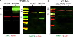
- Additional file 2: Figure S1. A, Western blot showing AMY3 transfected HEK293 cells demonstrate a marked increase in level of expression of CTR and RAMP3 proteins compared to wild-type (WT) HEK cells. B, in BV2 cells, RAMP3 protein expression shows a marked decreased after RAMP3 siRNA transfection compared to the control non-transfected cells. (JPEG 1495 kb)
- Submitted by
- Invitrogen Antibodies (provider)
- Main image
- Experimental details
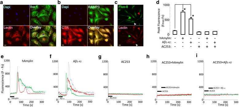
- Fig. 1 Microglial cells express functional amylin receptors. a Primary cultures of human fetal microglia (HFMs) that are stained with microglial antibody, Iba-1 (green), and DyLight-594-labeled lectin (red). b These primary cultured HFMs were also stained for the two dimeric proteins that are components of the amylin receptor 3 (AMY3) subtype, the calcitonin receptor (CTR), and the receptor-associated membrane protein 3 (RAMP3). c Cultured HFMs were loaded with the fluorescent intracellular calcium dye, Fluo-8L-AM (green), and with lectin (red), an in vivo microglial marker. Arrowheads indicate cells from which Ca 2+ signals were recorded. The same cell culture is also stained with lectin (red). The field in c shows that a majority of cells are microglia. d Summary of data on intracellular calcium changes after human amylin (hAmylin) and Abeta 1-42 without and with application of the amylin receptor antagonist, AC253 in HFM (* p < 0.05, n = 123 cells in 12 culture wells from four different batch of the culture cells). The candidate traces for intracellular calcium changes are illustrated in e - i . Elevations of Ca 2+ induced by acute (30 s) application of either hAmylin (1 muM, e ) or Abeta 1-42 (1 muM, f ). The changes in Ca 2+ were blocked by AC253 (10 muM, g - i ). Traces correspond to cells identified in c with arrowheads. Scale bar = 20 mum