Explore
 Validate
 Learn
Learn Flow cytometry
Antibody data
- Antibody Data
- Antigen structure
- References [8]
- Comments [0]
- Validations
- Flow cytometry [4]
- Other assay [1]
Submit
Validation data
Reference
Comment
Report error
- Product number
- 17-0819-42 - Provider product page

- Provider
- Invitrogen Antibodies
- Product name
- CD81 Monoclonal Antibody (1D6-CD81), APC, eBioscience™
- Antibody type
- Monoclonal
- Antigen
- Other
- Description
- Description: This 1D6-CD81 monoclonal antibody reacts with human CD81, which is also known as Target of Anti-Proliferative Ab-1 (TAPA-1). A member of the tetraspanin superfamily of transmembrane proteins, CD81 is widely expressed on immune cells such as B, T, and NK cells, monocytes, and eosinophils. Studies suggest that the highest expression of CD81 can be found on germinal center B cells. This protein can also be detected on non-Hodgkin lymphomas and diffuse large B-cell lymphomas. On B cells, CD81 exists in a complex with CD19, CD21, and Leu13. CD81 plays a role in segregating the CD19/CD21-B cell receptor complexes to lipid rafts to activate signal transduction. Finally, CD81 is a receptor for hepatitis C virus. The 1D6 monoclonal antibody has been reported to induce adhesion and reduce cell proliferation. Applications Reported: This 1D6-CD81 antibody has been reported for use in flow cytometric analysis. Applications Tested: This 1D6-CD81 antibody has been pre-titrated and tested by flow cytometric analysis of normal human peripheral blood cells. This can be used at 5 µL (1 µg) per test. A test is defined as the amount (µg) of antibody that will stain a cell sample in a final volume of 100 µL. Cell number should be determined empirically but can range from 10^5 to 10^8 cells/test. Excitation: 633-647 nm; Emission: 660 nm; Laser: Red Laser. Filtration: 0.2 µm post-manufacturing filtered.
- Reactivity
- Human
- Host
- Mouse
- Isotype
- IgG
- Antibody clone number
- 1D6-CD81
- Vial size
- 100 Tests
- Concentration
- 5 μL/Test
- Storage
- 4°C, store in dark, DO NOT FREEZE!
Submitted references uPAR(+) extracellular vesicles: a robust biomarker of resistance to checkpoint inhibitor immunotherapy in metastatic melanoma patients.
Open conformation of tetraspanins shapes interaction partner networks on cell membranes.
Comprehensive Cell Surface Antigen Analysis Identifies Transferrin Receptor Protein-1 (CD71) as a Negative Selection Marker for Human Neuronal Cells.
Continuous signaling of CD79b and CD19 is required for the fitness of Burkitt lymphoma B cells.
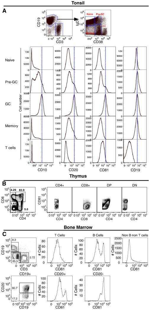
CD81 protein is expressed at high levels in normal germinal center B cells and in subtypes of human lymphomas.
CD81 gene defect in humans disrupts CD19 complex formation and leads to antibody deficiency.
CD81 (TAPA-1): a molecule involved in signal transduction and cell adhesion in the immune system.
The TAPA-1 molecule is associated on the surface of B cells with HLA-DR molecules.
Porcelli L, Guida M, De Summa S, Di Fonte R, De Risi I, Garofoli M, Caputo M, Negri A, Strippoli S, Serratì S, Azzariti A
Journal for immunotherapy of cancer 2021 May;9(5)
Journal for immunotherapy of cancer 2021 May;9(5)
Open conformation of tetraspanins shapes interaction partner networks on cell membranes.
Yang Y, Liu XR, Greenberg ZJ, Zhou F, He P, Fan L, Liu S, Shen G, Egawa T, Gross ML, Schuettpelz LG, Li W
The EMBO journal 2020 Sep 15;39(18):e105246
The EMBO journal 2020 Sep 15;39(18):e105246
Comprehensive Cell Surface Antigen Analysis Identifies Transferrin Receptor Protein-1 (CD71) as a Negative Selection Marker for Human Neuronal Cells.
Menon V, Thomas R, Elgueta C, Horl M, Osborn T, Hallett PJ, Bartos M, Isacson O, Pruszak J
Stem cells (Dayton, Ohio) 2019 Oct;37(10):1293-1306
Stem cells (Dayton, Ohio) 2019 Oct;37(10):1293-1306
Continuous signaling of CD79b and CD19 is required for the fitness of Burkitt lymphoma B cells.
He X, Kläsener K, Iype JM, Becker M, Maity PC, Cavallari M, Nielsen PJ, Yang J, Reth M
The EMBO journal 2018 Jun 1;37(11)
The EMBO journal 2018 Jun 1;37(11)
CD81 protein is expressed at high levels in normal germinal center B cells and in subtypes of human lymphomas.
Luo RF, Zhao S, Tibshirani R, Myklebust JH, Sanyal M, Fernandez R, Gratzinger D, Marinelli RJ, Lu ZS, Wong A, Levy R, Levy S, Natkunam Y
Human pathology 2010 Feb;41(2):271-80
Human pathology 2010 Feb;41(2):271-80
CD81 gene defect in humans disrupts CD19 complex formation and leads to antibody deficiency.
van Zelm MC, Smet J, Adams B, Mascart F, Schandené L, Janssen F, Ferster A, Kuo CC, Levy S, van Dongen JJ, van der Burg M
The Journal of clinical investigation 2010 Apr;120(4):1265-74
The Journal of clinical investigation 2010 Apr;120(4):1265-74
CD81 (TAPA-1): a molecule involved in signal transduction and cell adhesion in the immune system.
Levy S, Todd SC, Maecker HT
Annual review of immunology 1998;16:89-109
Annual review of immunology 1998;16:89-109
The TAPA-1 molecule is associated on the surface of B cells with HLA-DR molecules.
Schick MR, Levy S
Journal of immunology (Baltimore, Md. : 1950) 1993 Oct 15;151(8):4090-7
Journal of immunology (Baltimore, Md. : 1950) 1993 Oct 15;151(8):4090-7
No comments: Submit comment
Supportive validation
- Submitted by
- Invitrogen Antibodies (provider)
- Main image
- Experimental details
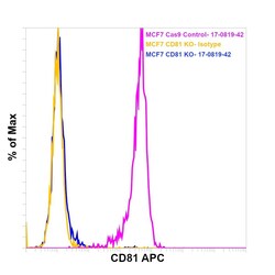
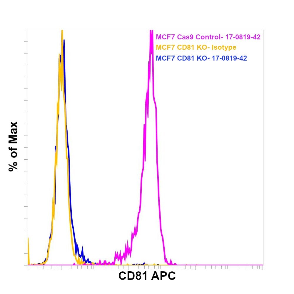
- Knockout of CD81 was achieved by CRISPR-Cas9 genome editing using LentiArray™ Lentiviral sgRNA (Product # A32042, Assay ID CRISPR661098_LV) and LentiArray Cas9 Lentivirus (Product # A32064). Flow cytometry analysis of CD81 was performed by staining THP-1 CD81 Knock out cells with 1 µg Mouse IgG1 kappa Isotype Control (P3.6.2.8.1), APC, eBioscience™ (Product # 17-4714-82, yellow histogram) or 1 µg CD81 Monoclonal Antibody (1D6-CD81), APC, eBioscience™ (Product # 17-0819-42, blue histogram). THP-1 Cas9 control cells were also stained with1 µg CD81 Monoclonal Antibody (1D6-CD81), APC, eBioscience™ (Product # 17-0819-42, pink histogram). Lossof signal was observed in the KOcells stained with CD81 antibody clone 1D6-CD81 but not in the control Cas9cells. Viable cells were used for analysis, as determined by Fixable Viability Dye eFluor™ 450 (Product # 65-0863-14).
- Submitted by
- Invitrogen Antibodies (provider)
- Main image
- Experimental details
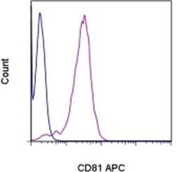
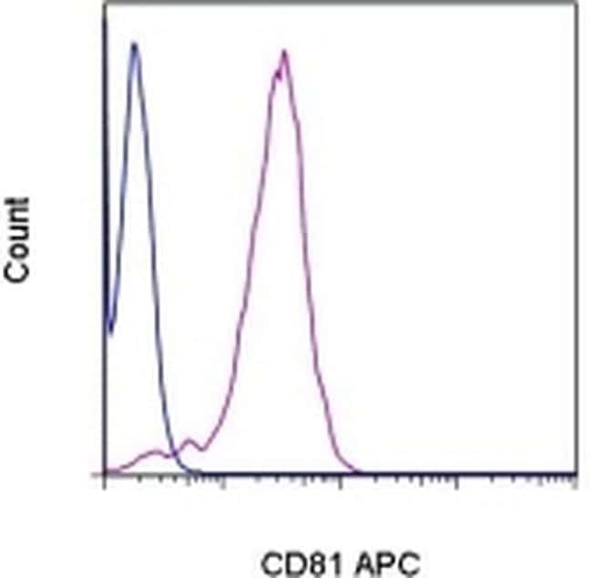
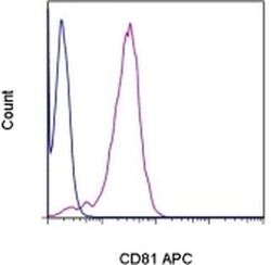
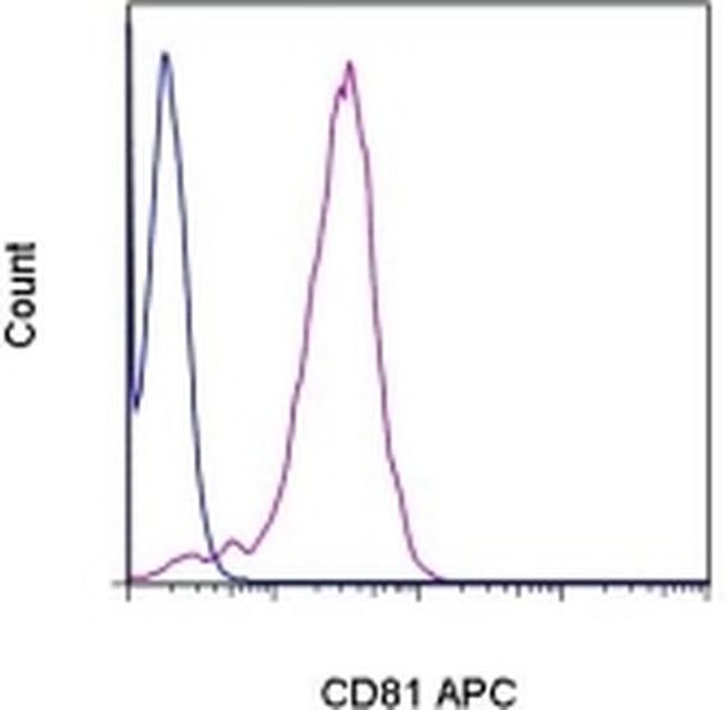
- Staining of normal human peripheral blood cells with Mouse IgG1 K Isotype Control APC (Product # 17-4714-81) (blue histogram) or Anti-Human CD81 APC (purple histogram). Cells in the lymphocyte gate were used for analysis.
- Submitted by
- Invitrogen Antibodies (provider)
- Main image

- Experimental details
- Knockout of CD81 was achieved by CRISPR-Cas9 genome editing using LentiArray™ Lentiviral sgRNA (Product # A32042, Assay ID CRISPR661098_LV) and LentiArray Cas9 Lentivirus (Product # A32064). Flow cytometry analysis of CD81 was performed by staining THP-1 CD81 Knock out cells with 1 µg Mouse IgG1 kappa Isotype Control (P3.6.2.8.1), APC, eBioscience™ (Product # 17-4714-82, yellow histogram) or 1 µg CD81 Monoclonal Antibody (1D6-CD81), APC, eBioscience™ (Product # 17-0819-42, blue histogram). THP-1 Cas9 control cells were also stained with1 µg CD81 Monoclonal Antibody (1D6-CD81), APC, eBioscience™ (Product # 17-0819-42, pink histogram). Lossof signal was observed in the KOcells stained with CD81 antibody clone 1D6-CD81 but not in the control Cas9cells. Viable cells were used for analysis, as determined by Fixable Viability Dye eFluor™ 450 (Product # 65-0863-14).

- Submitted by
- Invitrogen Antibodies (provider)
- Main image
- Experimental details
- Staining of normal human peripheral blood cells with Mouse IgG1 K Isotype Control APC (Product # 17-4714-81) (blue histogram) or Anti-Human CD81 APC (purple histogram). Cells in the lymphocyte gate were used for analysis.
Supportive validation
- Submitted by
- Invitrogen Antibodies (provider)
- Main image
- Experimental details
- NULL