Explore
 Validate
 Learn
Learn Western blot
 Flow cytometry
Antibody data
- Antibody Data
- Antigen structure
- References [4]
- Comments [0]
- Validations
- Flow cytometry [1]
Submit
Validation data
Reference
Comment
Report error
- Product number
- A01281-2 - Provider product page

- Provider
- Boster Biological Technology
- Product name
- Anti-TAPA1/CD81 Antibody Picoband™
- Antibody type
- Polyclonal
- Description
- Polyclonal antibody for CD81 detection. Host: Rabbit.Size: 100μg/vial. Tested applications: ELISA. Reactive species: Human. CD81 information: Subcellular Localization: Basolateral cell membrane; Tissue Specificity: Hematolymphoid, neuroectodermal and mesenchymal tumor cell lines.
- Reactivity
- Human, Mouse, Rat
- Host
- Rabbit
- Vial size
- 100μg/vial
- Concentration
- 0.5-1mg/ml, actual concentration vary by lot. Use suggested dilution ratio to decide dilution procedure.
- Storage
- At -20°C for one year. After reconstitution, at 4°C for one month. It can also be aliquoted and stored frozen at -20°C for a longer time. Avoid repeated freezing and thawing.
- Handling
- Add 0.2ml of distilled water will yield a concentration of 500ug/ml.
Submitted references Mechanobiological responses of astrocytes in optic nerve head due to biaxial stretch.
Exosomes from artesunate-treated bone marrow-derived mesenchymal stem cells transferring SNHG7 to promote osteogenesis via TAF15-RUNX2 pathway.
β-catenin-controlled tubular cell-derived exosomes play a key role in fibroblast activation via the OPN-CD44 axis.
Exosomal Surface Protein Detection with Quantum Dots and Immunomagnetic Capture for Cancer Detection.
Li Z, Peng F, Liu Z, Li S, Li L, Qian X
BMC ophthalmology 2022 Sep 16;22(1):368
BMC ophthalmology 2022 Sep 16;22(1):368
Exosomes from artesunate-treated bone marrow-derived mesenchymal stem cells transferring SNHG7 to promote osteogenesis via TAF15-RUNX2 pathway.
Huang MZ, Chen HY, Peng GX, Sun H, Peng HC, Li HY, Liu XH, Li Q
Regenerative medicine 2022 Nov;17(11):819-833
Regenerative medicine 2022 Nov;17(11):819-833
β-catenin-controlled tubular cell-derived exosomes play a key role in fibroblast activation via the OPN-CD44 axis.
Chen S, Zhang M, Li J, Huang J, Zhou S, Hou X, Ye H, Liu X, Xiang S, Shen W, Miao J, Hou FF, Liu Y, Zhou L
Journal of extracellular vesicles 2022 Mar;11(3):e12203
Journal of extracellular vesicles 2022 Mar;11(3):e12203
Exosomal Surface Protein Detection with Quantum Dots and Immunomagnetic Capture for Cancer Detection.
Vinduska V, Gallops CE, O'Connor R, Wang Y, Huang X
Nanomaterials (Basel, Switzerland) 2021 Jul 18;11(7)
Nanomaterials (Basel, Switzerland) 2021 Jul 18;11(7)
No comments: Submit comment
Supportive validation
- Submitted by
- Boster Biological Technology (provider)
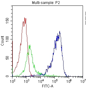
- Main image

- Experimental details
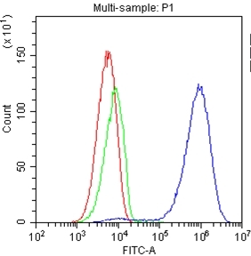
- Flow Cytometry analysis of PBMC cells using anti-TAPA1 antibody (A01281-2). Overlay histogram showing PBMC cells stained with A01281-2 (Blue line).The cells were blocked with 10% normal goat serum. And then incubated with rabbit anti-TAPA1 Antibody (A01281-2,1μg/1x106 cells) for 30 min at 20°C. DyLight?488 conjugated goat anti-rabbit IgG (BA1127, 5-10μg/1x106 cells) was used as secondary antibody for 30 minutes at 20°C. Isotype control antibody (Green line) was rabbit IgG (1μg/1x106) used under the same conditions. Unlabelled sample (Red line) was also used as a control.
- Additional image