Explore
 Validate
 Learn
Learn Western blot
 Immunocytochemistry
Antibody data
- Antibody Data
- Antigen structure
- References [6]
- Comments [0]
- Validations
- Immunocytochemistry [10]
- Immunohistochemistry [3]
- Flow cytometry [2]
- Other assay [5]
Submit
Validation data
Reference
Comment
Report error
- Product number
- MA5-32333 - Provider product page

- Provider
- Invitrogen Antibodies
- Product name
- CD81 Recombinant Rabbit Monoclonal Antibody (SN206-01)
- Antibody type
- Monoclonal
- Antigen
- Synthetic peptide
- Description
- Recombinant rabbit monoclonal antibodies are produced using in vitro expression systems. The expression systems are developed by cloning in the specific antibody DNA sequences from immunoreactive rabbits. Then, individual clones are screened to select the best candidates for production. The advantages of using recombinant rabbit monoclonal antibodies include: better specificity and sensitivity, lot-to-lot consistency, animal origin-free formulations, and broader immunoreactivity to diverse targets due to larger rabbit immune repertoire.
- Reactivity
- Human, Mouse, Rat
- Host
- Rabbit
- Isotype
- IgG
- Antibody clone number
- SN206-01
- Vial size
- 100 μL
- Concentration
- 1 mg/mL
- Storage
- Store at 4°C short term. For long term storage, store at -20°C, avoiding freeze/thaw cycles.
Submitted references Extracellular Vesicle-Encapsulated MicroRNA-375 from Bone Marrow-Derived Mesenchymal Stem Cells Inhibits Hepatocellular Carcinoma Progression through Regulating HOXB3-Mediated Wnt/β-Catenin Pathway.
Neural stem cell-derived exosomes suppress neuronal cell apoptosis by activating autophagy via miR-374-5p/STK-4 axis in spinal cord injury.
Ageing related thyroid deficiency increases brain-targeted transport of liver-derived ApoE4-laden exosomes leading to cognitive impairment.
Platelet membrane and stem cell exosome hybrid enhances cellular uptake and targeting to heart injury.
Macrophage secretion of miR-106b-5p causes renin-dependent hypertension.
The protective effects of MSC-EXO against pulmonary hypertension through regulating Wnt5a/BMP signalling pathway.
Yu Z, Liu J, Fan Q, Yu J, Ren X, Wang X
Analytical cellular pathology (Amsterdam) 2022;2022:9302496
Analytical cellular pathology (Amsterdam) 2022;2022:9302496
Neural stem cell-derived exosomes suppress neuronal cell apoptosis by activating autophagy via miR-374-5p/STK-4 axis in spinal cord injury.
Zhang L, Han P
Journal of musculoskeletal & neuronal interactions 2022 Sep 1;22(3):411-421
Journal of musculoskeletal & neuronal interactions 2022 Sep 1;22(3):411-421
Ageing related thyroid deficiency increases brain-targeted transport of liver-derived ApoE4-laden exosomes leading to cognitive impairment.
Zhang M, Gong W, Zhang D, Ji M, Chen B, Chen B, Li X, Zhou Y, Dong C, Wen G, Zhan X, Wu X, Cui L, Feng Y, Wang S, Yuan H, Xu E, Xia M, Verkhratsky A, Li B
Cell death & disease 2022 Apr 25;13(4):406
Cell death & disease 2022 Apr 25;13(4):406
Platelet membrane and stem cell exosome hybrid enhances cellular uptake and targeting to heart injury.
Hu S, Wang X, Li Z, Zhu D, Cores J, Wang Z, Li J, Mei X, Cheng X, Su T, Cheng K
Nano today 2021 Aug;39
Nano today 2021 Aug;39
Macrophage secretion of miR-106b-5p causes renin-dependent hypertension.
Oh J, Matkovich SJ, Riek AE, Bindom SM, Shao JS, Head RD, Barve RA, Sands MS, Carmeliet G, Osei-Owusu P, Knutsen RH, Zhang H, Blumer KJ, Nichols CG, Mecham RP, Baldán Á, Benitez BA, Sequeira-Lopez ML, Gomez RA, Bernal-Mizrachi C
Nature communications 2020 Sep 23;11(1):4798
Nature communications 2020 Sep 23;11(1):4798
The protective effects of MSC-EXO against pulmonary hypertension through regulating Wnt5a/BMP signalling pathway.
Zhang Z, Ge L, Zhang S, Wang J, Jiang W, Xin Q, Luan Y
Journal of cellular and molecular medicine 2020 Dec;24(23):13938-13948
Journal of cellular and molecular medicine 2020 Dec;24(23):13938-13948
No comments: Submit comment
Supportive validation
- Submitted by
- Invitrogen Antibodies (provider)
- Main image
- Experimental details
- Immunocytochemical analysis of CD81 in 293 cells using a CD81 Monoclonal antibody (Product # MA5-32333) as seen in green. The nuclear counter stain is DAPI (blue). Cells were fixed in paraformaldehyde, permeabilised with 0.25% Triton X100/PBS.

- Submitted by
- Invitrogen Antibodies (provider)
- Main image

- Experimental details
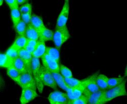
- Immunocytochemical analysis of CD81 in A549 cells using a CD81 Monoclonal antibody (Product # MA5-32333) as seen in green. The nuclear counter stain is DAPI (blue). Cells were fixed in paraformaldehyde, permeabilised with 0.25% Triton X100/PBS.

- Submitted by
- Invitrogen Antibodies (provider)
- Main image

- Experimental details
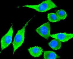
- Immunocytochemical analysis of CD81 in SH-SY-5Y cells using a CD81 Monoclonal antibody (Product # MA5-32333) as seen in green. The nuclear counter stain is DAPI (blue). Cells were fixed in paraformaldehyde, permeabilised with 0.25% Triton X100/PBS.

- Submitted by
- Invitrogen Antibodies (provider)
- Main image

- Experimental details
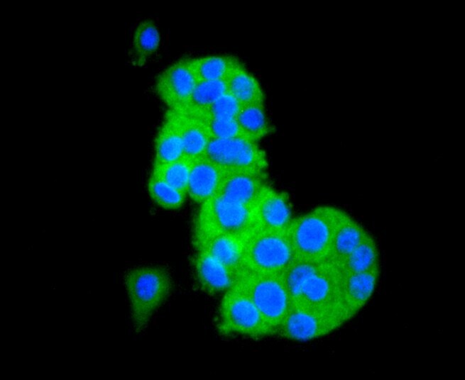
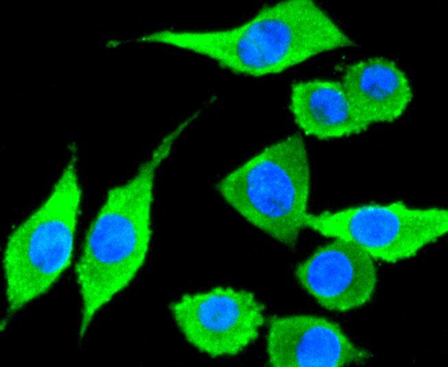
- Immunocytochemical analysis of CD81 in PC-12 cells using a CD81 Monoclonal antibody (Product # MA5-32333) as seen in green. The nuclear counter stain is DAPI (blue). Cells were fixed in paraformaldehyde, permeabilised with 0.25% Triton X100/PBS.
- Submitted by
- Invitrogen Antibodies (provider)
- Main image

- Experimental details
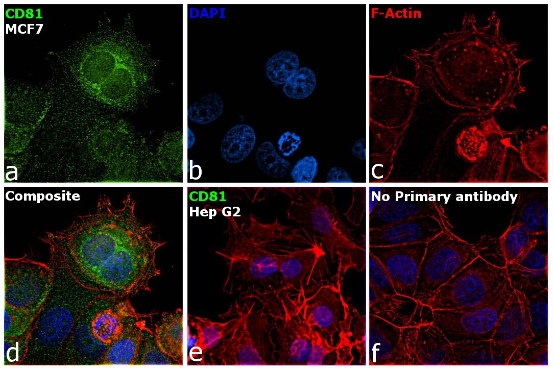
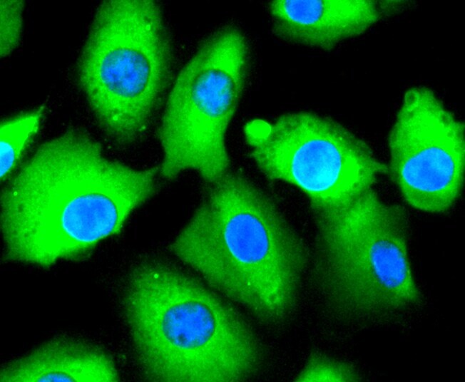
- Immunofluorescence analysis of CD81 antigen was performed using 70% confluent log phase MCF7 cells. The cells were fixed with 4% paraformaldehyde for 5 minutes, permeabilized with 0.1% Triton™ X-100 for 15 minutes, and blocked with 2% BSA for overnight at room temperature. The cells were labeled with CD81 Recombinant Rabbit Monoclonal Antibody (SN206-01) (Product # MA5-32333) at 1:100 dilution in 0.1% BSA, incubated at 4 degree celsius overnight and then labeled with Donkey anti-Rabbit IgG (H+L) Highly Cross-Adsorbed Secondary Antibody, Alexa Fluor Plus 488 (Product # A32790), (1:2000 dilution), for 45 minutes at room temperature (Panel a: Green). Nuclei (Panel b: Blue) were stained with ProLong™ Diamond Antifade Mountant with DAPI (Product # P36962). F-actin (Panel c: Red) was stained with Rhodamine Phalloidin (Product # R415, 1:300). Panel d represents the merged image showing peroxisomes, vesicles, ER, membrane localization. Panel e represents low expressing model, Hep G2. Panel f represents control cells with no primary antibody to assess background. The images were captured at 60X magnification.
- Submitted by
- Invitrogen Antibodies (provider)
- Main image

- Experimental details
- Immunocytochemical analysis of CD81 in 293 cells using a CD81 Monoclonal antibody (Product # MA5-32333) as seen in green. The nuclear counter stain is DAPI (blue). Cells were fixed in paraformaldehyde, permeabilised with 0.25% Triton X100/PBS.

- Submitted by
- Invitrogen Antibodies (provider)
- Main image

- Experimental details
- Immunocytochemical analysis of CD81 in A549 cells using a CD81 Monoclonal antibody (Product # MA5-32333) as seen in green. The nuclear counter stain is DAPI (blue). Cells were fixed in paraformaldehyde, permeabilised with 0.25% Triton X100/PBS.
- Submitted by
- Invitrogen Antibodies (provider)
- Main image
- Experimental details
- Immunocytochemical analysis of CD81 in SH-SY-5Y cells using a CD81 Monoclonal antibody (Product # MA5-32333) as seen in green. The nuclear counter stain is DAPI (blue). Cells were fixed in paraformaldehyde, permeabilised with 0.25% Triton X100/PBS.
- Submitted by
- Invitrogen Antibodies (provider)
- Main image

- Experimental details
- Immunocytochemical analysis of CD81 in PC-12 cells using a CD81 Monoclonal antibody (Product # MA5-32333) as seen in green. The nuclear counter stain is DAPI (blue). Cells were fixed in paraformaldehyde, permeabilised with 0.25% Triton X100/PBS.

- Submitted by
- Invitrogen Antibodies (provider)
- Main image

- Experimental details
- Immunofluorescence analysis of CD81 antigen was performed using 70% confluent log phase MCF7 cells. The cells were fixed with 4% paraformaldehyde for 5 minutes, permeabilized with 0.1% Triton™ X-100 for 15 minutes, and blocked with 2% BSA for overnight at room temperature. The cells were labeled with CD81 Recombinant Rabbit Monoclonal Antibody (SN206-01) (Product # MA5-32333) at 1:100 dilution in 0.1% BSA, incubated at 4 degree celsius overnight and then labeled with Donkey anti-Rabbit IgG (H+L) Highly Cross-Adsorbed Secondary Antibody, Alexa Fluor Plus 488 (Product # A32790), (1:2000 dilution), for 45 minutes at room temperature (Panel a: Green). Nuclei (Panel b: Blue) were stained with ProLong™ Diamond Antifade Mountant with DAPI (Product # P36962). F-actin (Panel c: Red) was stained with Rhodamine Phalloidin (Product # R415, 1:300). Panel d represents the merged image showing peroxisomes, vesicles, ER, membrane localization. Panel e represents low expressing model, Hep G2. Panel f represents control cells with no primary antibody to assess background. The images were captured at 60X magnification.

Supportive validation
- Submitted by
- Invitrogen Antibodies (provider)
- Main image

- Experimental details
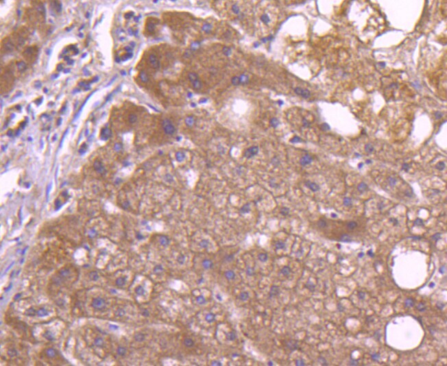
- Immunohistochemical analysis of CD81 of paraffin-embedded Human liver tissue using a CD81 Monoclonal antibody (Product #MA5-32333). Counter stained with hematoxylin.
- Submitted by
- Invitrogen Antibodies (provider)
- Main image
- Experimental details
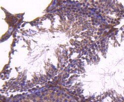
- Immunohistochemical analysis of CD81 of paraffin-embedded Mouse testis tissue using a CD81 Monoclonal antibody (Product #MA5-32333). Counter stained with hematoxylin.

- Submitted by
- Invitrogen Antibodies (provider)
- Main image
- Experimental details
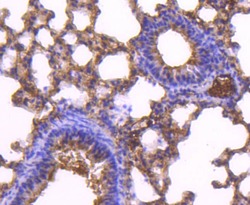
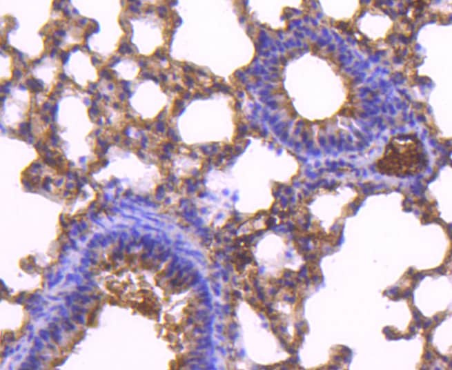
- Immunohistochemical analysis of CD81 of paraffin-embedded Mouse lung tissue using a CD81 Monoclonal antibody (Product #MA5-32333). Counter stained with hematoxylin.
Supportive validation
- Submitted by
- Invitrogen Antibodies (provider)
- Main image
- Experimental details
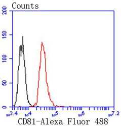
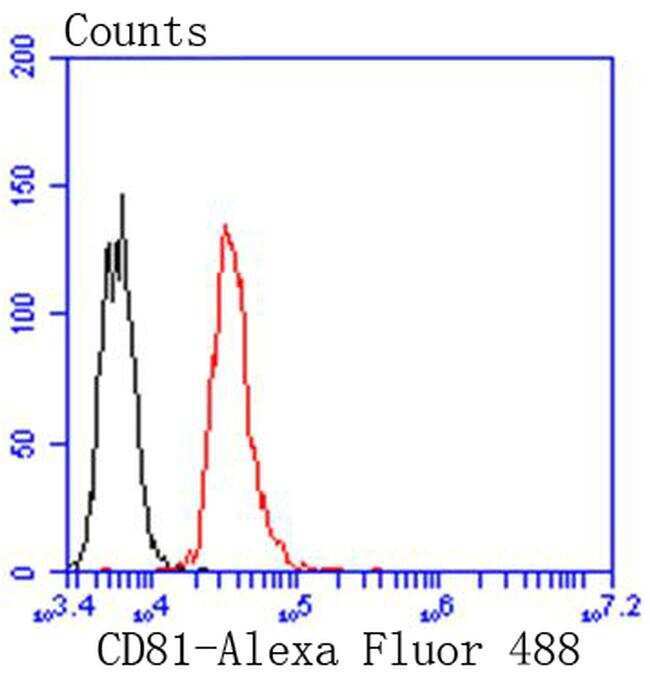
- Flow Cytometric analysis of CD81 in Jurkat cells using a CD81 Monoclonal Antibody (Product # MA5-32333) at a dilution of 1:50, as seen in red compared with an unlabelled control (cells without incubation with primary antibody; black). Alexa Fluor 488-conjugated goat anti rabbit IgG was used as the secondary antibody.
- Submitted by
- Invitrogen Antibodies (provider)
- Main image

- Experimental details
- Flow Cytometric analysis of CD81 in Jurkat cells using a CD81 Monoclonal Antibody (Product # MA5-32333) at a dilution of 1:50, as seen in red compared with an unlabelled control (cells without incubation with primary antibody; black). Alexa Fluor 488-conjugated goat anti rabbit IgG was used as the secondary antibody.

Supportive validation
- Submitted by
- Invitrogen Antibodies (provider)
- Main image
- Experimental details
- NULL
- Submitted by
- Invitrogen Antibodies (provider)
- Main image
- Experimental details
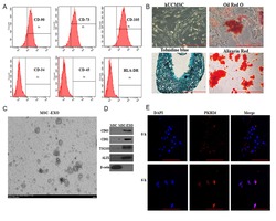
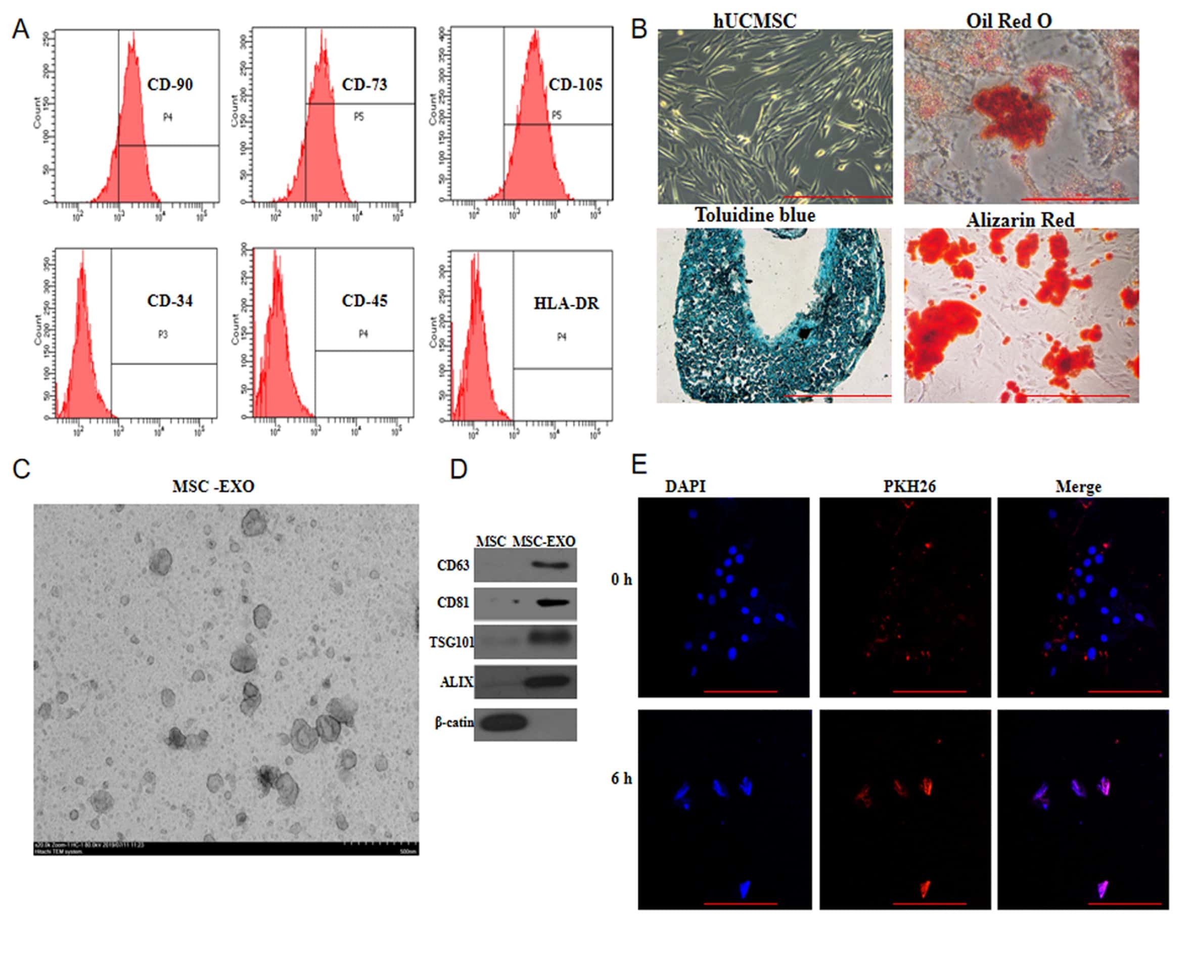
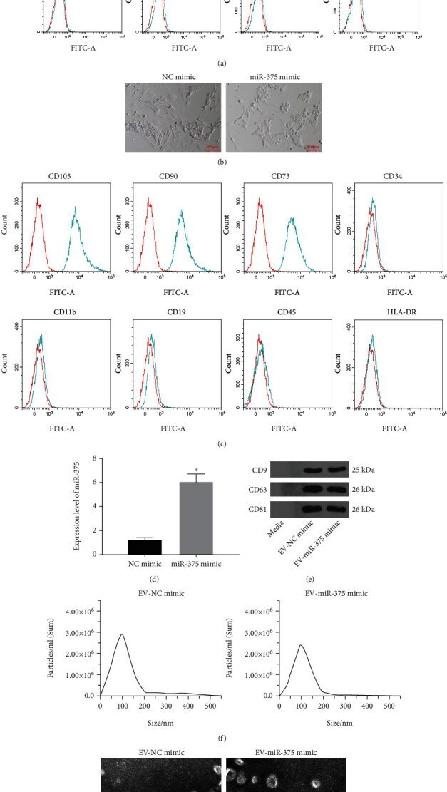
- Figure 2 EV from miR-375 overexpression-modified BM-MSCs are successfully extracted. (a) Expressions of BM-MSC markers CD73, CD90, and CD105 and hematopoietic and endothelial cell markers CD34, CD11b, CD19, CD45, and HLA-DR were measured by flow cytometry. (b). Morphological observation of BM-MSCs. (c) Expression of BM-MSC markers CD73, CD90, and CD105 and hematopoietic and endothelial cell markers CD34, CD11b, CD19, CD45, and HLA-DR in BM-MSCs transfected with NC mimic and miR-375 mimic were measured by flow cytometry. (d) miR-375 expression in BM-MSCs after transfection using RT-qPCR. (e) Expression of EV markers CD9, CD63, and CD81 was determined by Western blot assay. (f) The size distribution of EV analyzed using NTA. (g) TEM observation of EV structure. * p < 0.05 vs. the NC mimic group. All data were expressed as mean +- standard deviation of at least three independent experiments. Data analysis between the two groups was performed by unpaired t -test.
- Submitted by
- Invitrogen Antibodies (provider)
- Main image
- Experimental details
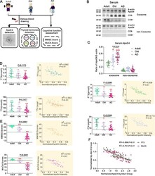
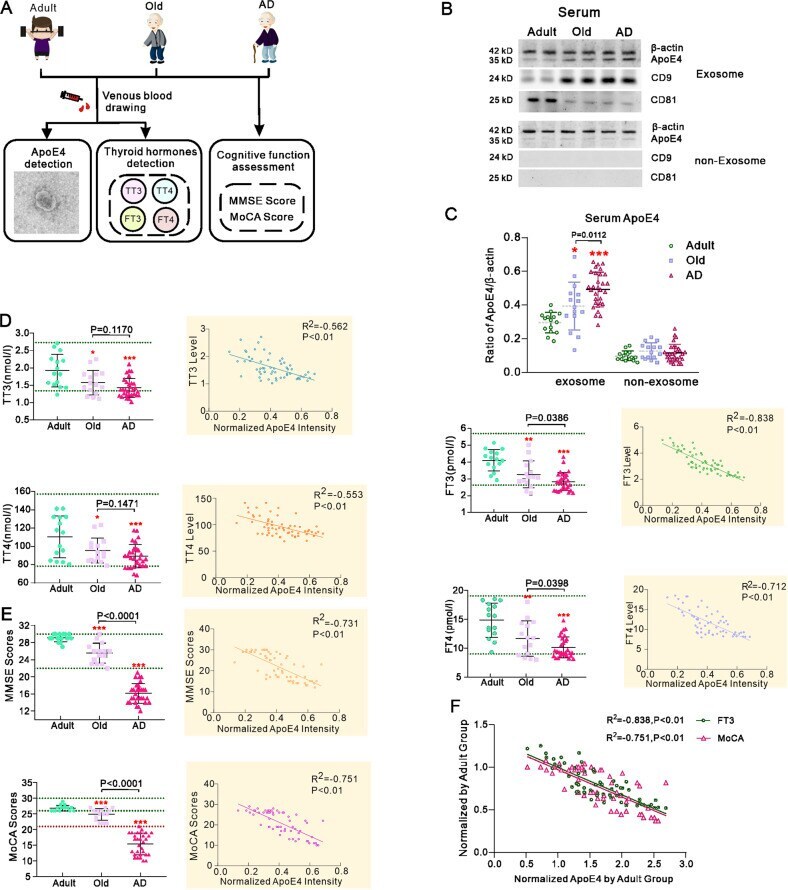
- Correlation of ApoE4 level in serum exosomes with thyroid hormones levels and cognitive performance in human subjects. A Enroled 60 carriers of ApoE4 allele included 15 adult healthy subjects, 15 old healthy subjects and 30 old AD patients. Collected serum was used to extract exosomes and measure TT3, FT3, TT4 and FT4. Participants cognitive abilities were assessed by MMSE and MoCA scoring. B Representative western blots of ApoE4 and exosomal specific marker CD81 and CD9 in the serum extracted exosomes and the supernatant from which exosomes precipitate was removed after ultracentrifuge. C Normalised intensities of ApoE4 by beta-actin. D TT3, FT3, TT4 and FT4 in adult, old and AD groups. Correlation analyses between the ApoE4 level in serum exosomes and TH levels are shown in inserts at the right. E MMSE and MoCA scores; correlation analyses between the ApoE4 level in serum exosomes and these scores are shown in inserts at the right. F Multiple correlation analysis of the normalised ApoE4 in serum exosomes with the normalised MoCA scores and serum FT3 in adult group. The individual measured value was plotted and shown as mean +- SD. One-way ANOVA for comparisons including more than two groups; unpaired two-tailed t -test for two-group comparisons. * p < 0.05, ** p < 0.01, *** p < 0.001.
- Submitted by
- Invitrogen Antibodies (provider)
- Main image
- Experimental details
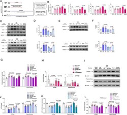
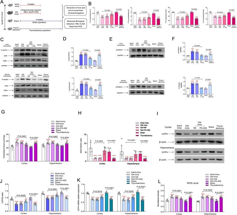
- The effects of L-thy and ApoE4 inhibitor on the exosome transport of h-ApoE4 and cerebral oxidative stress in ApoE4-KI mice. A Old mice were randomly intraperitoneally injected with normal saline (NS), 20 mug/kg/day L-thy or 200 mg/kg/day ApoE4 inhibitor PH-002 for 14 days; 2 months old mice randomly underwent thyroidectomy or sham surgery. Four weeks after surgery measurements were made. B TT3, FT3, TT4 and FT4 values are plotted as mean +- SD, n = 6. One-way ANOVA for comparisons including more than two groups; unpaired two-tailed t -test for two-group comparisons. As comparison with adult group, * p < 0.05, ** p < 0.01, *** p < 0.001. C Representative Western blots of h-ApoE4 in the exosomes extracted from liver and serum, CD9 and CD81 were used as the specific markers of exosomes. D Expression ratio of h-ApoE4 and GAPDH are plotted as mean +- SD, n = 6. E Representative Western blots of ATP6AP1 in the exosomes extracted from liver and serum. F Ratio of ATP6AP1 normalised to GAPDH is plotted as mean +- SD, n = 6. One-way ANOVA for comparisons including more than two groups; unpaired two-tailed t -test for two-group comparisons. As comparison with adult group, * p < 0.05, ** p < 0.01, *** p < 0.001. G , H Cholesterol and the ratio of GSH/GSSG in cortex and hippocampus measured by ELISA are plotted as mean +- SD, n = 6. I Representative Western blots of UCP4 in cortex and in hippocampus. J Ratio of UCP4 to beta-actin is plotted as mean +- SD, n = 6. K mRNA expression of UCP4
- Submitted by
- Invitrogen Antibodies (provider)
- Main image
- Experimental details
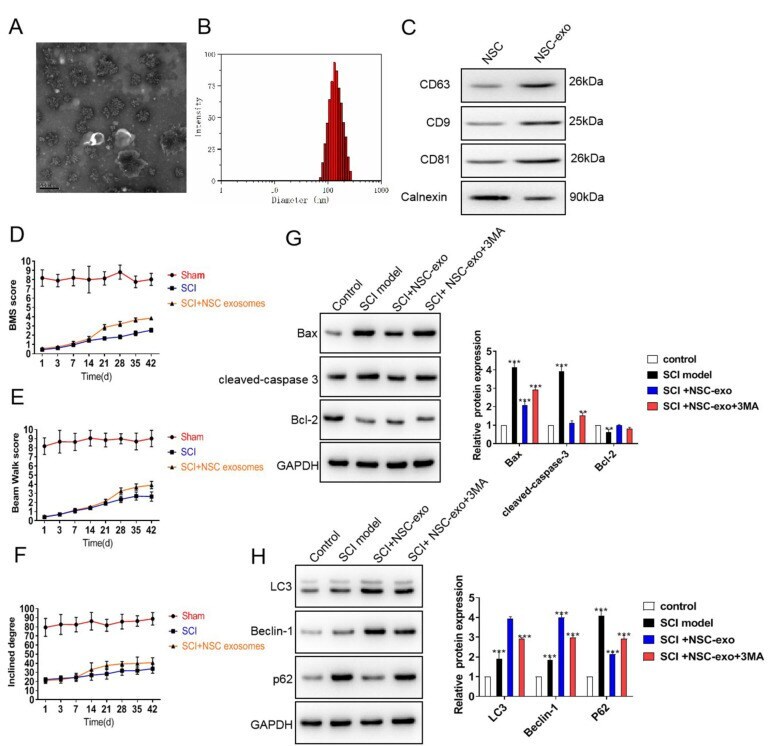
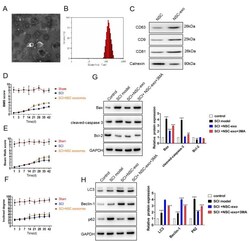
- Neural stem cell (NSC)-derived exosome could protect against spinal cord injury (SCI). Exosomes were isolated and confirmed by transmission electron microscopy (TEM) (A), NTA (B), and western blot of exosome markers (C). (D-F) Behavioral assessment by BMS, beam walking, and inclined plane testing after SCI. (G and H) Western blot analysis for apoptosis markers and autophagy flux markers, respectively. The band intensities were analyzed using the gray values in Image J software.