Explore
 Validate
 Learn
Learn Western blot
 Flow cytometry
Antibody data
- Antibody Data
- Antigen structure
- References [7]
- Comments [0]
- Validations
- Flow cytometry [9]
Submit
Validation data
Reference
Comment
Report error
- Product number
- NBP1-44861 - Provider product page

- Provider
- Novus Biologicals
- Proper citation
- Novus Cat#NBP1-44861, RRID:AB_10008097
- Product name
- Mouse Monoclonal CD81 Antibody
- Antibody type
- Monoclonal
- Description
- Protein A purified. The antibody M38 reacts with CD81, a 25 kDa member of the tetraspanin family, expressed on majority of cells.
- Reactivity
- Human, Feline, Rabbit
- Host
- Mouse
- Isotype
- IgG
- Vial size
- 0.1 mg
- Concentration
- 1 mg/ml
- Storage
- Store at 4C. Do not freeze.
Submitted references Slow Release of HIV-1 Protein Nef from Vesicle-like Structures Is Inhibited by Cytosolic Calcium Elevation in Single Human Microglia.
Selective enrichment of tetraspan proteins on the internal vesicles of multivesicular endosomes and on exosomes secreted by human B-lymphocytes.
Selective enrichment of tetraspan proteins on the internal vesicles of multivesicular endosomes and on exosomes secreted by human B-lymphocytes.
Molecular analyses of the association of CD4 with two members of the transmembrane 4 superfamily, CD81 and CD82.
C33 antigen and M38 antigen recognized by monoclonal antibodies inhibitory to syncytium formation by human T cell leukemia virus type 1 are both members of the transmembrane 4 superfamily and associate with each other and with CD4 or CD8 in T cells.
Identification of membrane antigen C33 recognized by monoclonal antibodies inhibitory to human T-cell leukemia virus type 1 (HTLV-1)-induced syncytium formation: altered glycosylation of C33 antigen in HTLV-1-positive T cells.
Identification of membrane antigen C33 recognized by monoclonal antibodies inhibitory to human T-cell leukemia virus type 1 (HTLV-1)-induced syncytium formation: altered glycosylation of C33 antigen in HTLV-1-positive T cells.
Stenovec M, Lasič E, Dominkuš PP, Bobnar ST, Zorec R, Lenassi M, Kreft M
Molecular neurobiology 2019 Jan;56(1):102-118
Molecular neurobiology 2019 Jan;56(1):102-118
Selective enrichment of tetraspan proteins on the internal vesicles of multivesicular endosomes and on exosomes secreted by human B-lymphocytes.
Escola JM, Kleijmeer MJ, Stoorvogel W, Griffith JM, Yoshie O, Geuze HJ
The Journal of biological chemistry 1998 Aug 7;273(32):20121-7
The Journal of biological chemistry 1998 Aug 7;273(32):20121-7
Molecular analyses of the association of CD4 with two members of the transmembrane 4 superfamily, CD81 and CD82.
Imai T, Kakizaki M, Nishimura M, Yoshie O
Journal of immunology (Baltimore, Md. : 1950) 1995 Aug 1;155(3):1229-39
Journal of immunology (Baltimore, Md. : 1950) 1995 Aug 1;155(3):1229-39
C33 antigen and M38 antigen recognized by monoclonal antibodies inhibitory to syncytium formation by human T cell leukemia virus type 1 are both members of the transmembrane 4 superfamily and associate with each other and with CD4 or CD8 in T cells.
Imai T, Yoshie O
Journal of immunology (Baltimore, Md. : 1950) 1993 Dec 1;151(11):6470-81
Journal of immunology (Baltimore, Md. : 1950) 1993 Dec 1;151(11):6470-81
Identification of membrane antigen C33 recognized by monoclonal antibodies inhibitory to human T-cell leukemia virus type 1 (HTLV-1)-induced syncytium formation: altered glycosylation of C33 antigen in HTLV-1-positive T cells.
Fukudome K, Furuse M, Imai T, Nishimura M, Takagi S, Hinuma Y, Yoshie O
Journal of virology 1992 Mar;66(3):1394-401
Journal of virology 1992 Mar;66(3):1394-401
No comments: Submit comment
Supportive validation
- Submitted by
- Novus Biologicals (provider)
- Main image
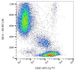
- Experimental details
- Flow Cytometry: CD81 Antibody (M38) [NBP1-44861] - Surface staining of CD81 in human peripheral blood with anti-CD81 (M38) APC-CyTM7.

- Submitted by
- Novus Biologicals (provider)
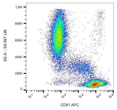
- Main image

- Experimental details
- Flow Cytometry: CD81 Antibody (M38) [NBP1-44861] - Human peripheral blood with anti-CD81 (M38) APC.
- Submitted by
- Novus Biologicals (provider)
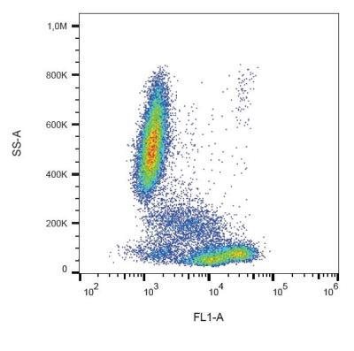
- Main image

- Experimental details
- Flow Cytometry: CD81 Antibody (M38) [NBP1-44861] - Human peripheral blood with anti-CD81 (M38) FITC.
- Submitted by
- Novus Biologicals (provider)
- Main image

- Experimental details
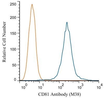
- Flow Cytometry: CD81 Antibody (M38) [NBP1-44861] - Flow Cytometry: CD81 Antibody (M38) [PE] [NBP1-44861PE] - A cell surface stain was performed on THP-1 cells with CD81 antibody (M38) NBP1-44861 (blue) and a matched isotype control NBP2-27287 (orange). Cells were incubated in an antibody dilution of 1:100 for 20 minutes at room temperature. Both antibodies were conjugated to PE. Image using the PE form of this antibody.
- Submitted by
- Novus Biologicals (provider)
- Main image

- Experimental details
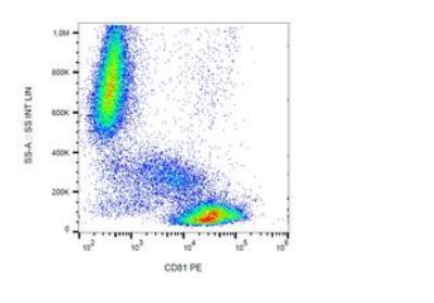
- Flow Cytometry: CD81 Antibody (M38) [NBP1-44861] - Staining of CD81 in human peripheral blood with anti-CD81 (M38) PE.
- Submitted by
- Novus Biologicals (provider)
- Main image
- Experimental details
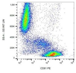
- Flow Cytometry: CD81 Antibody (M38) [NBP1-44861] - Staining of CD81 in human peripheral blood with anti-CD81 (M38) PE.

- Submitted by
- Novus Biologicals (provider)
- Main image
- Experimental details
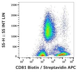
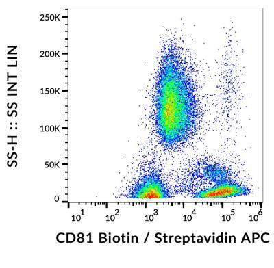
- Flow Cytometry: CD81 Antibody (M38) [NBP1-44861] - Staining of CD81 in human peripheral blood with anti-CD81 (M38) biotin, streptavidin-APC.
- Submitted by
- Novus Biologicals (provider)
- Main image
- Experimental details
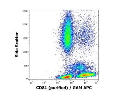
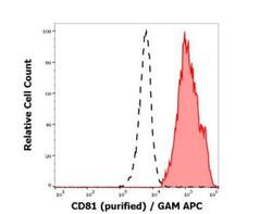
- Flow Cytometry: CD81 Antibody (M38) [NBP1-44861] - Separation of human lymphocytes (red-filled) from neutrophil granulocytes (black-dashed) in flow cytometry analysis (surface staining) of human peripheral whole blood stained using anti-human CD81 (M38) purified antibody (concentration in sample 4 ug/ml) GAM APC.

- Submitted by
- Novus Biologicals (provider)
- Main image
- Experimental details
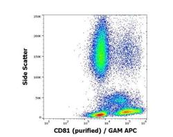
- Flow Cytometry: CD81 Antibody (M38) [NBP1-44861] - Surface staining pattern of human peripheral blood stained using anti-human CD81 (M38) purified antibody (concentration in sample 4 ug/ml) GAM APC.