Explore
 Validate
 Learn
Learn Western blot
Antibody data
- Antibody Data
- Antigen structure
- References [1]
- Comments [0]
- Validations
- Western blot [4]
- Immunocytochemistry [1]
Submit
Validation data
Reference
Comment
Report error
- Product number
- PA5-29759 - Provider product page

- Provider
- Invitrogen Antibodies
- Product name
- CRALBP Polyclonal Antibody
- Antibody type
- Polyclonal
- Antigen
- Recombinant protein fragment
- Description
- Recommended positive controls: HepG2, mouse eye. Predicted reactivity: Mouse (91%), Rat (91%), Xenopus laevis (82%), Chicken (86%), Bovine (92%). Store product as a concentrated solution. Centrifuge briefly prior to opening the vial.
- Reactivity
- Human, Mouse
- Host
- Rabbit
- Isotype
- IgG
- Vial size
- 100 µL
- Concentration
- 1.41 mg/mL
- Storage
- Store at 4°C short term. For long term storage, store at -20°C, avoiding freeze/thaw cycles.
Submitted references Stem Cell Derived Retinal Pigment Epithelium: The Role of Pigmentation as Maturation Marker and Gene Expression Profile Comparison with Human Endogenous Retinal Pigment Epithelium.
Bennis A, Jacobs JG, Catsburg LAE, Ten Brink JB, Koster C, Schlingemann RO, van Meurs J, Gorgels TGMF, Moerland PD, Heine VM, Bergen AA
Stem cell reviews and reports 2017 Oct;13(5):659-669
Stem cell reviews and reports 2017 Oct;13(5):659-669
No comments: Submit comment
Supportive validation
- Submitted by
- Invitrogen Antibodies (provider)
- Main image
- Experimental details
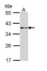
- Western blot analysis of CRALBP using 30 µg of HepG2 lysate. Samples were loaded onto a 10% SDS-PAGE gel and probed with a CRALBP polyclonal antibody (Product # PA5-29759) at a dilution of 1:3000.
- Submitted by
- Invitrogen Antibodies (provider)
- Main image
- Experimental details
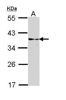
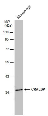
- Western Blot analysis of CRALBP was performed by separating 50 µg of mouse tissue extract by 10% SDS-PAGE. Proteins were transferred to a membrane and probed with a CRALBP Polyclonal Antibody (Product # PA5-29759) at a dilution of 1:1000.
- Submitted by
- Invitrogen Antibodies (provider)
- Main image
- Experimental details
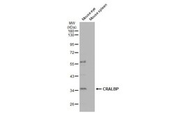
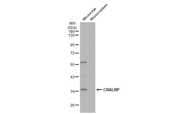
- Western Blot using CRALBP Polyclonal Antibody (Product # PA5-29759). Various tissue extracts (50 µg) were separated by 10% SDS-PAGE, and the membrane was blotted with CRALBP Polyclonal Antibody (Product # PA5-29759) diluted at 1:1,000. The HRP-conjugated anti-rabbit IgG antibody was used to detect the primary antibody, and the signal was developed with Trident ECL plus-Enhanced.
- Submitted by
- Invitrogen Antibodies (provider)
- Main image
- Experimental details
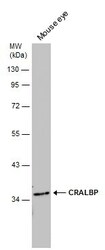
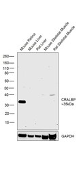
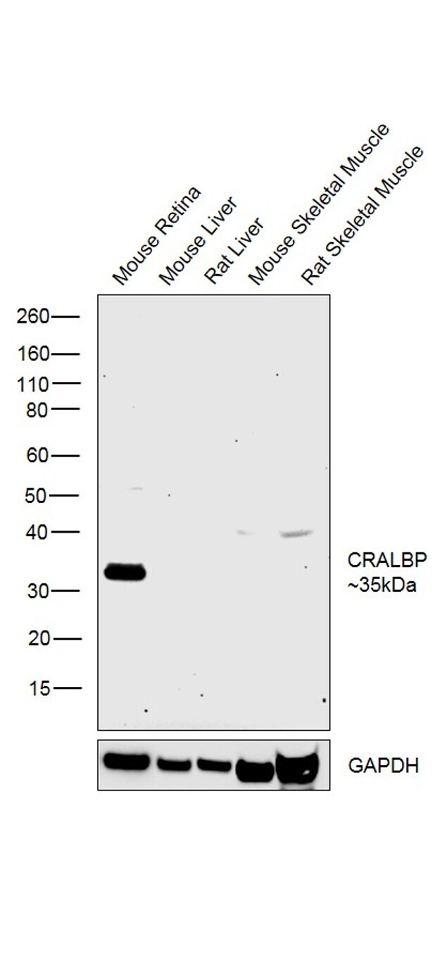
- Western blot was performed using Anti-CRALBP Rabbit Polyclonal Antibody (Product # PA5-29759) and a 35kDa band corresponding to CRALBP was observed across tissues tested except Mouse Liver, Rat Liver, Mouse Skeletal Muscle and Rat Skeletal Muscle which are reported negative for CRALBP expression. Tissue extracts (30 µg lysate) of Mouse Retina (Lane 1), Mouse Liver (Lane 2), Rat Liver (Lane 3), Mouse Skeletal Muscle (Lane 4) and Rat Skeletal Muscle (Lane 5) were electrophoresed using Novex® NuPAGE® 4-12 % Bis-Tris gel (Product # NP0322BOX). Resolved proteins were then transferred onto a nitrocellulose membrane (Product # IB23001) by iBlot® 2 Dry Blotting System (Product # IB21001). The blot was probed with the primary antibody (1:3000 dilution) and detected by chemiluminescence Goat Anti-Rabbit IgG Secondary Antibody, HRP conjugate (Product # A27036, 1:4000 dilution) using the iBright FL 1000 (Product # A32752). Chemiluminescent detection was performed using Novex® ECL Chemiluminescent Substrate Reagent Kit (Product # WP20005).
Supportive validation
- Submitted by
- Invitrogen Antibodies (provider)
- Main image
- Experimental details
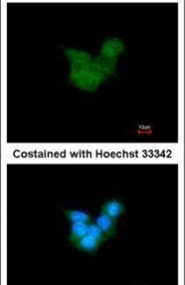
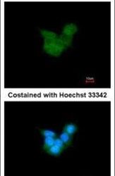
- Immunofluorescent analysis of CRALBP in methanol-fixed HepG2 cells using a CRALBP polyclonal antibody (Product # PA5-29759) at a 1:200 dilution.