Explore
 Validate
 Learn
Learn Western blot
 Immunocytochemistry
Antibody data
- Antibody Data
- Antigen structure
- References [5]
- Comments [0]
- Validations
- Immunocytochemistry [2]
- Immunoprecipitation [1]
- Immunohistochemistry [3]
- Other assay [2]
Submit
Validation data
Reference
Comment
Report error
- Product number
- PA5-21594 - Provider product page

- Provider
- Invitrogen Antibodies
- Product name
- MAD2 Polyclonal Antibody
- Antibody type
- Polyclonal
- Antigen
- Synthetic peptide
- Description
- Recommended positive controls: 293T, NCIN87, U2OS, HepG2, Molt4, Raji. Predicted reactivity: Rhesus Monkey (100%), Bovine (100%). Store product as a concentrated solution. Centrifuge briefly prior to opening the vial.
- Reactivity
- Human, Rat
- Host
- Rabbit
- Isotype
- IgG
- Vial size
- 100 μL
- Concentration
- 1.12 mg/mL
- Storage
- Store at 4°C short term. For long term storage, store at -20°C, avoiding freeze/thaw cycles.
Submitted references CCAR2 controls mitotic progression through spatiotemporal regulation of Aurora B.
Phosphorylation and Pin1 binding to the LIC1 subunit selectively regulate mitotic dynein functions.
Atypical APC/C-dependent degradation of Mcl-1 provides an apoptotic timer during mitotic arrest.
TC Mps1 12, a novel Mps1 inhibitor, suppresses the growth of hepatocellular carcinoma cells via the accumulation of chromosomal instability.
Polo-like kinase 1 inhibitor BI2536 causes mitotic catastrophe following activation of the spindle assembly checkpoint in non-small cell lung cancer cells.
Ryu J, Kim JE
Cell death & disease 2022 Jun 7;13(6):534
Cell death & disease 2022 Jun 7;13(6):534
Phosphorylation and Pin1 binding to the LIC1 subunit selectively regulate mitotic dynein functions.
Kumari A, Kumar C, Pergu R, Kumar M, Mahale SP, Wasnik N, Mylavarapu SVS
The Journal of cell biology 2021 Dec 6;220(12)
The Journal of cell biology 2021 Dec 6;220(12)
Atypical APC/C-dependent degradation of Mcl-1 provides an apoptotic timer during mitotic arrest.
Allan LA, Skowyra A, Rogers KI, Zeller D, Clarke PR
The EMBO journal 2018 Sep 3;37(17)
The EMBO journal 2018 Sep 3;37(17)
TC Mps1 12, a novel Mps1 inhibitor, suppresses the growth of hepatocellular carcinoma cells via the accumulation of chromosomal instability.
Choi M, Min YH, Pyo J, Lee CW, Jang CY, Kim JE
British journal of pharmacology 2017 Jun;174(12):1810-1825
British journal of pharmacology 2017 Jun;174(12):1810-1825
Polo-like kinase 1 inhibitor BI2536 causes mitotic catastrophe following activation of the spindle assembly checkpoint in non-small cell lung cancer cells.
Choi M, Kim W, Cheon MG, Lee CW, Kim JE
Cancer letters 2015 Feb 28;357(2):591-601
Cancer letters 2015 Feb 28;357(2):591-601
No comments: Submit comment
Supportive validation
- Submitted by
- Invitrogen Antibodies (provider)
- Main image

- Experimental details
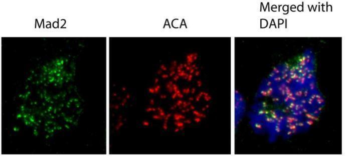
- Immunofluorescence analysis of human osteosarcoma cell line U2OS, using MAD2L1 antibody (Product # PA5-21594) at 1:1,000 dilution.

- Submitted by
- Invitrogen Antibodies (provider)
- Main image

- Experimental details
- Immunofluorescence analysis of human osteosarcoma cell line U2OS, using MAD2L1 antibody (Product # PA5-21594) at 1:1,000 dilution.
Supportive validation
- Submitted by
- Invitrogen Antibodies (provider)
- Main image

- Experimental details
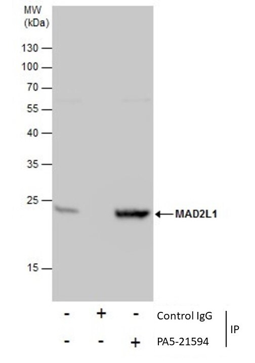
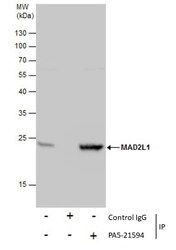
- Immunoprecipitation of MAD2L1 was performed in 293T whole cell extracts using 5 µg of MAD2 Polyclonal Antibody (Product # PA5-21594). Samples were transferred to a membrane and probed with MAD2 Polyclonal Antibody as a primary antibody and an HRP-conjugated anti-Rabbit IgG was used as a secondary antibody.
Supportive validation
- Submitted by
- Invitrogen Antibodies (provider)
- Main image
- Experimental details
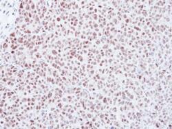
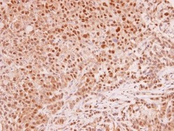
- Immunohistochemical analysis of paraffin-embedded DLD-1 xenograft, using Mad2L1 (Product # PA5-21594) antibody at 1:500 dilution. Antigen Retrieval: EDTA based buffer, pH 8.0, 15 min.
- Submitted by
- Invitrogen Antibodies (provider)
- Main image

- Experimental details
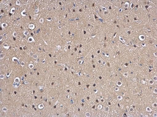
- MAD2 Polyclonal Antibody detects MAD2L1 protein at nucleus on rat fore brain by immunohistochemical analysis. Sample: Paraffin-embedded rat fore brain. MAD2 Polyclonal Antibody (Product # PA5-21594) dilution: 1:500. Antigen Retrieval: EDTA based buffer, pH 8.0, 15 min.
- Submitted by
- Invitrogen Antibodies (provider)
- Main image
- Experimental details
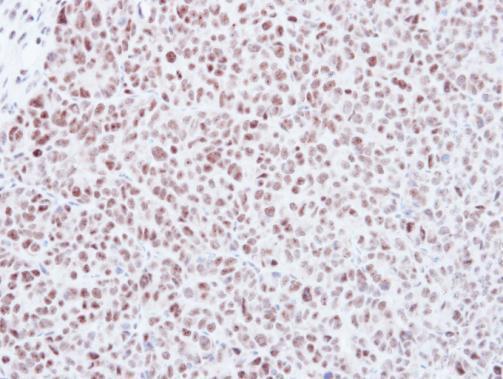
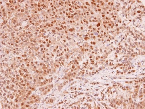
- Immunohistochemistry (Paraffin) analysis of MAD2 was performed in paraffin-embedded human A549 xenograft tissue using MAD2 Polyclonal Antibody (Product # PA5-21594) at a dilution of 1:250.
Supportive validation
- Submitted by
- Invitrogen Antibodies (provider)
- Main image
- Experimental details
- Immunoprecipitation of MAD2L1 was performed in 293T whole cell extracts using 5 µg of MAD2 Polyclonal Antibody (Product # PA5-21594). Samples were transferred to a membrane and probed with MAD2 Polyclonal Antibody as a primary antibody and an HRP-conjugated anti-Rabbit IgG was used as a secondary antibody.

- Submitted by
- Invitrogen Antibodies (provider)
- Main image
- Experimental details
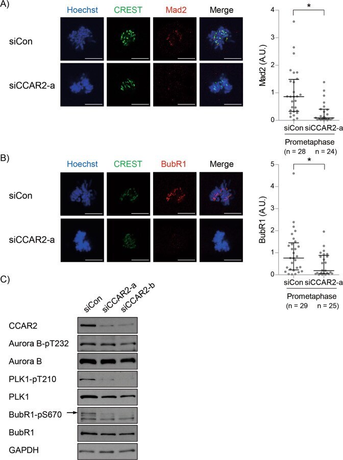
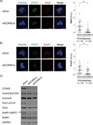
- Fig. 5 CCAR2 deficiency induces inactivation of spindle assembly checkpoint. A , B Asynchronous A549 cells were stained with Hoechst, CREST, and anti-Mad2 or anti-BubR1 antibodies. Fluorescence intensity of kinetochore-associated Mad2 (A) or BubR1 (B) in siCCAR2-a cells was normalized to that of siCon at prometaphase. The number of prometaphase in all experiments ( n ) was presented in each graph. The first and third bars are the 25th and 75th percentiles, respectively, and the second bar is the median. * p < 0.05, significantly different from corresponding siCon cells (Mann-Whitney U test); A.U., arbitrary units; scale bar, 10 mum. C Cells were arrested at the G1/S boundary by double thymidine block and then released for 9 h. MG132 (30 muM) was treated for the last three hours to block anaphase onset. The amount of each protein was determined by western blotting.