Explore
 Validate
 Learn
Learn Western blot
 Immunocytochemistry
 Immunohistochemistry
Antibody data
- Antibody Data
- Antigen structure
- References [4]
- Comments [0]
- Validations
- Immunocytochemistry [4]
- Other assay [2]
Submit
Validation data
Reference
Comment
Report error
- Product number
- PA3-750 - Provider product page

- Provider
- Invitrogen Antibodies
- Product name
- PRDX1 Polyclonal Antibody
- Antibody type
- Polyclonal
- Antigen
- Synthetic peptide
- Description
- PA3-750 detects Peroxiredoxin 1 protein in human, rat and mouse samples. This antibody is specific for Prx 1 and shows no cross-reactivity with other Prx isoforms. PA3-750 has successfully been used in Western blot and immunofluorescence procedures. By Western blot, this antibody detects an ~26 kDa protein representing Prx 1 in human PC3 cell lysate. The PA3-750 immunizing peptide corresponds to amino acid residues 103-114 from human Prx 1.
- Reactivity
- Human, Mouse, Rat
- Host
- Rabbit
- Isotype
- IgG
- Vial size
- 100 μL
- Concentration
- Conc. Not Determined
- Storage
- -20°C, Avoid Freeze/Thaw Cycles
Submitted references Mitochondrial CoQ deficiency is a common driver of mitochondrial oxidants and insulin resistance.
Proteomic analysis of the effects of aged garlic extract and its FruArg component on lipopolysaccharide-induced neuroinflammatory response in microglial cells.
Proteomic profiling identifies cyclooxygenase-2-independent global proteomic changes by celecoxib in colorectal cancer cells.
Nonredundant antioxidant defense by multiple two-cysteine peroxiredoxins in human prostate cancer cells.
Fazakerley DJ, Chaudhuri R, Yang P, Maghzal GJ, Thomas KC, Krycer JR, Humphrey SJ, Parker BL, Fisher-Wellman KH, Meoli CC, Hoffman NJ, Diskin C, Burchfield JG, Cowley MJ, Kaplan W, Modrusan Z, Kolumam G, Yang JY, Chen DL, Samocha-Bonet D, Greenfield JR, Hoehn KL, Stocker R, James DE
eLife 2018 Feb 6;7
eLife 2018 Feb 6;7
Proteomic analysis of the effects of aged garlic extract and its FruArg component on lipopolysaccharide-induced neuroinflammatory response in microglial cells.
Zhou H, Qu Z, Mossine VV, Nknolise DL, Li J, Chen Z, Cheng J, Greenlief CM, Mawhinney TP, Brown PN, Fritsche KL, Hannink M, Lubahn DB, Sun GY, Gu Z
PloS one 2014;9(11):e113531
PloS one 2014;9(11):e113531
Proteomic profiling identifies cyclooxygenase-2-independent global proteomic changes by celecoxib in colorectal cancer cells.
Lou J, Fatima N, Xiao Z, Stauffer S, Smythers G, Greenwald P, Ali IU
Cancer epidemiology, biomarkers & prevention : a publication of the American Association for Cancer Research, cosponsored by the American Society of Preventive Oncology 2006 Sep;15(9):1598-606
Cancer epidemiology, biomarkers & prevention : a publication of the American Association for Cancer Research, cosponsored by the American Society of Preventive Oncology 2006 Sep;15(9):1598-606
Nonredundant antioxidant defense by multiple two-cysteine peroxiredoxins in human prostate cancer cells.
Shen C, Nathan C
Molecular medicine (Cambridge, Mass.) 2002 Feb;8(2):95-102
Molecular medicine (Cambridge, Mass.) 2002 Feb;8(2):95-102
No comments: Submit comment
Supportive validation
- Submitted by
- Invitrogen Antibodies (provider)
- Main image
- Experimental details
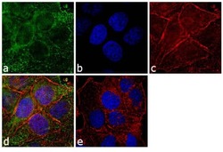
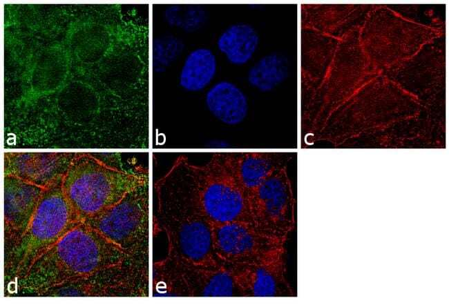
- Immunofluorescence analysis of Peroxiredoxin 1 was performed using 70% confluent log phase T-47D cells. The cells were fixed with 4% paraformaldehyde for 10 minutes, permeabilized with 0.1% Triton™ X-100 for 10 minutes, and blocked with 1% BSA for 1 hour at room temperature. The cells were labeled with PRDX1 Rabbit Polyclonal (Product # PA3-750) at 1:250 dilution in 0.1% BSA and incubated for 3 hours at room temperature and then labeled with Goat anti-Rabbit IgG (H+L) Superclonal™ Secondary Antibody, Alexa Fluor® 488 conjugate (Product # A27034) at a dilution of 1:2,000 for 45 minutes at room temperature (Panel a: green). Nuclei (Panel b: blue) were stained with SlowFade® Gold Antifade Mountant with DAPI (Product # S36938). F-actin (Panel c: red) was stained with Rhodamine Phalloidin (Product # R415, 1:300). Panel d represents the merged image showing cytosolic and nuclear localization. Panel e shows the no primary antibody control. The images were captured at 60X magnification.

- Submitted by
- Invitrogen Antibodies (provider)
- Main image
- Experimental details
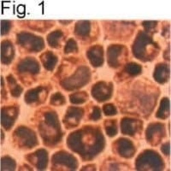
- Immunocytochemical staining of Prx 1 in TSU-Pr1 cells using Product # PA3-750.
- Submitted by
- Invitrogen Antibodies (provider)
- Main image

- Experimental details
- Immunofluorescence analysis of Peroxiredoxin 1 was performed using 70% confluent log phase T-47D cells. The cells were fixed with 4% paraformaldehyde for 10 minutes, permeabilized with 0.1% Triton™ X-100 for 10 minutes, and blocked with 1% BSA for 1 hour at room temperature. The cells were labeled with PRDX1 Rabbit Polyclonal (Product # PA3-750) at 1:250 dilution in 0.1% BSA and incubated for 3 hours at room temperature and then labeled with Goat anti-Rabbit IgG (Heavy Chain) Superclonal™ Secondary Antibody, Alexa Fluor® 488 conjugate (Product # A27034) at a dilution of 1:2,000 for 45 minutes at room temperature (Panel a: green). Nuclei (Panel b: blue) were stained with SlowFade® Gold Antifade Mountant with DAPI (Product # S36938). F-actin (Panel c: red) was stained with Rhodamine Phalloidin (Product # R415, 1:300). Panel d represents the merged image showing cytosolic and nuclear localization. Panel e shows the no primary antibody control. The images were captured at 60X magnification.
- Submitted by
- Invitrogen Antibodies (provider)
- Main image

- Experimental details
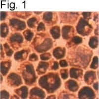
- Immunocytochemical staining of Prx 1 in TSU-Pr1 cells using Product # PA3-750.

Supportive validation
- Submitted by
- Invitrogen Antibodies (provider)
- Main image
- Experimental details
- NULL
- Submitted by
- Invitrogen Antibodies (provider)
- Main image

- Experimental details
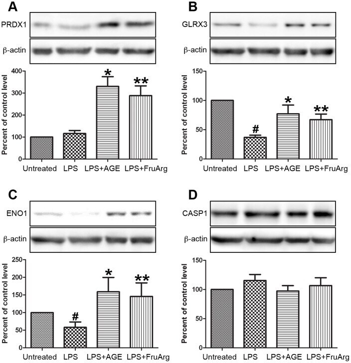
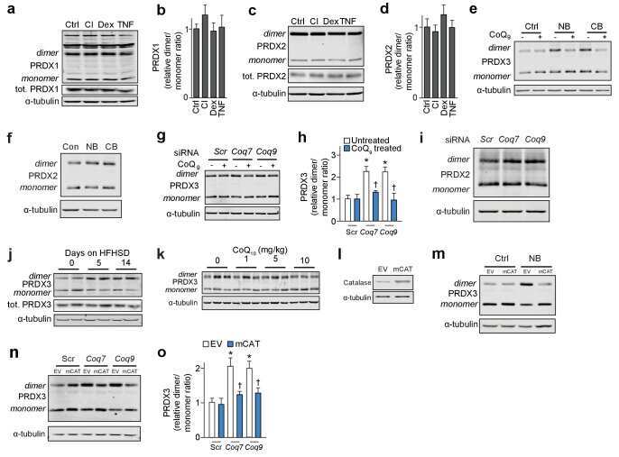
- Figure 5 Validation of expression profiling of proteins by Western blotting. Four of the identified proteins, PRDX1 ( A ), GLRX3 ( B ), ENO1 ( C ), and CASP1 ( D ), responding to AGE and/or FruArg treatment in LPS-stimulated BV-2 cells were validated using Western blotting. Protein intensities were quantified by Image J software, normalized to beta-actin, and expressed as percentage of untreated controls. Data are means +- SEM (n>=5); #, P