 Explore
 Validate
 Learn
Learn Western blot
Antibody data
- Antibody Data
- Antigen structure
- References [2]
- Comments [0]
- Validations
- Western blot [2]
Submit
Validation data
Reference
Comment
Report error
- Product number
- AF3488 - Provider product page

- Provider
- R&D Systems
- Product name
- Human/Mouse/Rat Peroxiredoxin 1 Antibody
- Antibody type
- Polyclonal
- Description
- Antigen Affinity-purified. Detects human, mouse and rat Peroxiredoxin 1 in Western blots. In direct ELISAs, less than 1% cross-reactivity with recombinant human Peroxiredoxin 3 or 4 is observed.
- Reactivity
- Human, Mouse, Rat
- Host
- Goat
- Conjugate
- Unconjugated
- Antigen sequence
Q06830- Isotype
- IgG
- Vial size
- 100 ug
- Concentration
- LYOPH
- Storage
- Use a manual defrost freezer and avoid repeated freeze-thaw cycles. 12 months from date of receipt, -20 to -70 °C as supplied. 1 month, 2 to 8 °C under sterile conditions after reconstitution. 6 months, -20 to -70 °C under sterile conditions after reconstitution.
Submitted references Electrophiles modulate glutathione reductase activity via alkylation and upregulation of glutathione biosynthesis.
The impact of tyrosine kinase 2 (Tyk2) on the proteome of murine macrophages and their response to lipopolysaccharide (LPS).
Jobbagy S, Vitturi DA, Salvatore SR, Turell L, Pires MF, Kansanen E, Batthyany C, Lancaster JR Jr, Freeman BA, Schopfer FJ
Redox biology 2019 Feb;21:101050
Redox biology 2019 Feb;21:101050
The impact of tyrosine kinase 2 (Tyk2) on the proteome of murine macrophages and their response to lipopolysaccharide (LPS).
Radwan M, Miller I, Grunert T, Marchetti-Deschmann M, Vogl C, O'Donoghue N, Dunn MJ, Kolbe T, Allmaier G, Gemeiner M, Müller M, Strobl B
Proteomics 2008 Sep;8(17):3469-85
Proteomics 2008 Sep;8(17):3469-85
No comments: Submit comment
Supportive validation
- Submitted by
- R&D Systems (provider)
- Main image
- Experimental details
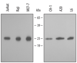
- Detection of Human/Mouse/Rat Peroxiredoxin 1 by Western Blot. Western blot shows lysates of Jurkat human acute T cell leukemia cell line, Raji human Burkitt's lymphoma cell line, MCF-7 human breast cancer cell line, CH-1 mouse B cell lymphoma cell line, A20 mouse B cell lymphoma cell line, and L6 rat myoblast cell line . PVDF membrane was probed with 0.2 µg/mL of Goat Anti-Human/Mouse/Rat Peroxiredoxin 1 Antigen Affinity-purified Polyclonal Antibody (Catalog # AF3488) followed by HRP-conjugated Anti-Goat IgG Secondary Antibody (Catalog # HAF109). A specific band was detected for Peroxiredoxin 1 at approximately 22 kDa (as indicated). This experiment was conducted using Immunoblot Buffer Group 2.

- Submitted by
- R&D Systems (provider)
- Main image
- Experimental details
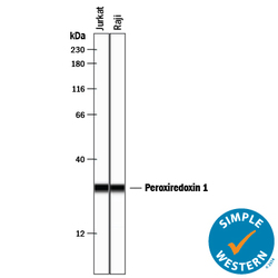
- Detection of Human Peroxiredoxin 1 by Simple WesternTM. Simple Western lane view shows lysates of Jurkat human acute T cell leukemia cell line and Raji human Burkitt's lymphoma cell line, loaded at 0.2 mg/mL. A specific band was detected for Peroxiredoxin 1 at approximately 29 kDa (as indicated) using 2 µg/mL of Goat Anti-Human/Mouse/Rat Peroxiredoxin 1 Antigen Affinity-purified Polyclonal Antibody (Catalog # AF3488) followed by 1:50 dilution of HRP-conjugated Anti-Goat IgG Secondary Antibody (Catalog # HAF109). This experiment was conducted under reducing conditions and using the 12-230 kDa separation system.
