Explore
 Validate
 Learn
Learn Western blot
 ELISA
ELISA Immunocytochemistry
Antibody data
- Antibody Data
- Antigen structure
- References [1]
- Comments [0]
- Validations
- Immunocytochemistry [2]
- Other assay [1]
Submit
Validation data
Reference
Comment
Report error
- Product number
- PA5-112712 - Provider product page

- Provider
- Invitrogen Antibodies
- Product name
- MCT2 Polyclonal Antibody
- Antibody type
- Polyclonal
- Antigen
- Recombinant full-length protein
- Reactivity
- Human
- Host
- Rabbit
- Isotype
- IgG
- Vial size
- 100 μg
- Concentration
- 1.7 mg/mL
- Storage
- -20°C or -80°C if preferred
Submitted references Landscape of surfaceome and endocytome in human glioma is divergent and depends on cellular spatial organization.
Governa V, Talbot H, Gonçalves de Oliveira K, Cerezo-Magaña M, Bång-Rudenstam A, Johansson MC, Månsson AS, Forsberg-Nilsson K, Marko-Varga G, Enríquez Pérez J, Darabi A, Malmström J, Bengzon J, Welinder C, Belting M
Proceedings of the National Academy of Sciences of the United States of America 2022 Mar 1;119(9)
Proceedings of the National Academy of Sciences of the United States of America 2022 Mar 1;119(9)
No comments: Submit comment
Supportive validation
- Submitted by
- Invitrogen Antibodies (provider)
- Main image
- Experimental details
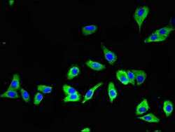
- Immunocytochemichal analysis of MCT2 in HepG2 cells using a Polyclonal antibody (Product # PA5-112712) at a dilution of 1:100. An Alexa Fluor 488-congugated Goat Anti-Rabbit IgG(H+L) secondary antibdoy was used.

- Submitted by
- Invitrogen Antibodies (provider)
- Main image
- Experimental details
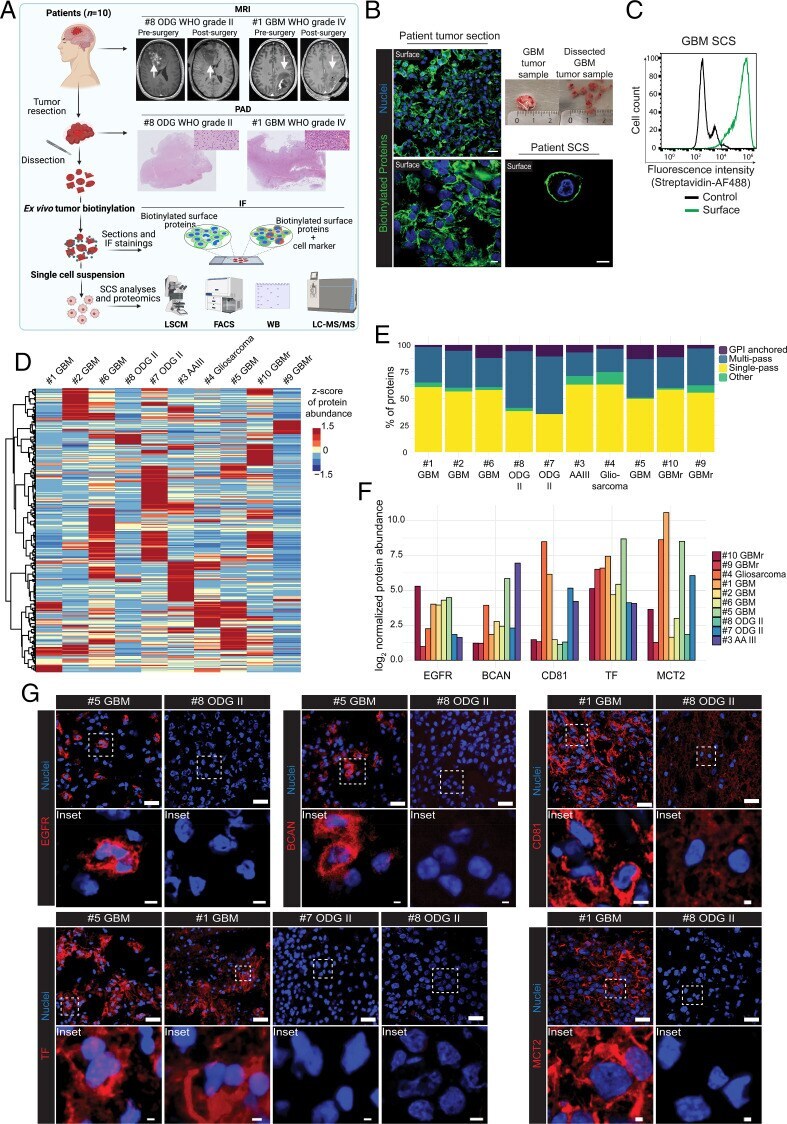
- Immunocytochemistry analysis of MCT2 in Hela cells. The sample was incubated with a MCT2 polyclonal antibody (Product # PA5-112712) at a dilution of 1:200, followed by Alexa Fluor 488-congugated AffiniPure Goat Anti-Rabbit IgG(H+L).

Supportive validation
- Submitted by
- Invitrogen Antibodies (provider)
- Main image
- Experimental details
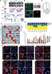
- Fig. 3. TS-MAP uncovers a divergent surfaceome landscape in patient gliomas. ( A ) TS-MAP was applied in a mixed cohort ( n = 10) of patient gliomas. Shown are MRI (presurgery and postsurgery within 48 h) and histology for pathological-anatomical diagnostics (PAD) according to clinical routine, here exemplified by patient #8ODG with a low-grade ODG and #1GBM representing a high-grade GBM. Freshly resected tumors were biotinylated ex vivo for downstream analyses, as indicated. ( B ) Confocal microscopy shows biotinylation (green) of intact patient tumor. Higher magnification ( Bottom ) indicates surface labeling, which is further supported by Airyscan imaging of disintegrated SCS ( Bottom Right ), showing specific plasma membrane labeling. Top Right shows example of fresh tumor dissection into pieces of 0.3 to 0.5 cm in diameter prior to biotinylation (ruler scale in centimeters). (Scale bars, 20 mum [ Top Left ], 5 mum [ Bottom Left and Right ].) ( C ) FACS quantification of streptavidin-AF488 association with nonbiotinylated (control) and biotinylated (surface) in a representative GBM patient tissue SCS. ( D ) TS-MAP was applied in a pilot cohort of 10 freshly resected patient tumors, including low-grade ODG (WHO grade II), high-grade anaplastic astrocytoma (AA, WHO grade III), primary glioblastoma (GBM, WHO grade IV), recurrent GBM (GBMr), and gliosarcoma (WHO grade IV). Heatmap of SURFME protein abundance demonstrates divergent expression profile among patient tumors. ( E