Explore
 Validate
 Learn
Learn Western blot
 Immunocytochemistry
Antibody data
- Antibody Data
- Antigen structure
- References [1]
- Comments [0]
- Validations
- Immunocytochemistry [3]
- Immunohistochemistry [6]
Submit
Validation data
Reference
Comment
Report error
- Product number
- PA5-54580 - Provider product page

- Provider
- Invitrogen Antibodies
- Product name
- ACADS Polyclonal Antibody
- Antibody type
- Polyclonal
- Antigen
- Recombinant protein fragment
- Description
- Immunogen sequence: ELPETHQMLL QTCRDFAEKE LFPIAAQVDK EHLFPAAQVK KMGGLGLLAM DVPEELGGAG LDYLAYAIAM EEISRGCAST GVIMSVNNSL YLGPILKFGS KEQKQAWVTP FTSGDKIGCF ALSEPGNGS Highest antigen sequence identity to the following orthologs: Mouse - 88%, Rat - 89%.
- Reactivity
- Human, Mouse, Rat
- Host
- Rabbit
- Isotype
- IgG
- Vial size
- 100 μL
- Concentration
- 0.3 mg/mL
- Storage
- Store at 4°C short term. For long term storage, store at -20°C, avoiding freeze/thaw cycles.
Submitted references Induction of LEF1 by MYC activates the WNT pathway and maintains cell proliferation.
Hao YH, Lafita-Navarro MC, Zacharias L, Borenstein-Auerbach N, Kim M, Barnes S, Kim J, Shay J, DeBerardinis RJ, Conacci-Sorrell M
Cell communication and signaling : CCS 2019 Oct 17;17(1):129
Cell communication and signaling : CCS 2019 Oct 17;17(1):129
No comments: Submit comment
Supportive validation
- Submitted by
- Invitrogen Antibodies (provider)
- Main image
- Experimental details
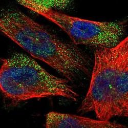
- Immunofluorescent staining of ACADS in human cell line U-251 MG shows positivity in cytoplasm, centrosome & nucleus but excluded from the nucleoli. Samples were probed using an ACADS Polyclonal Antibody (Product # PA5-54580).
- Submitted by
- Invitrogen Antibodies (provider)
- Main image

- Experimental details
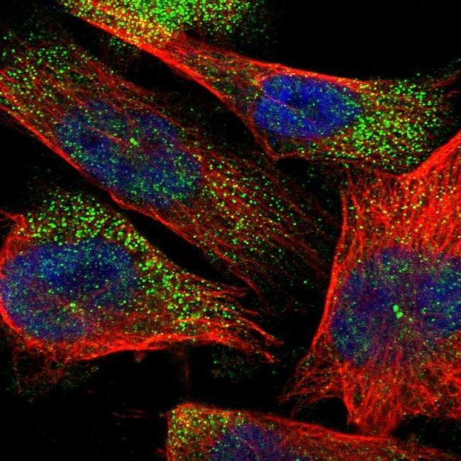
- Immunofluorescent staining of ACADS in human cell line Hep G2 using ACADS Polyclonal Antibody (Product # PA5-54580) shows localization to mitochondria.

- Submitted by
- Invitrogen Antibodies (provider)
- Main image

- Experimental details
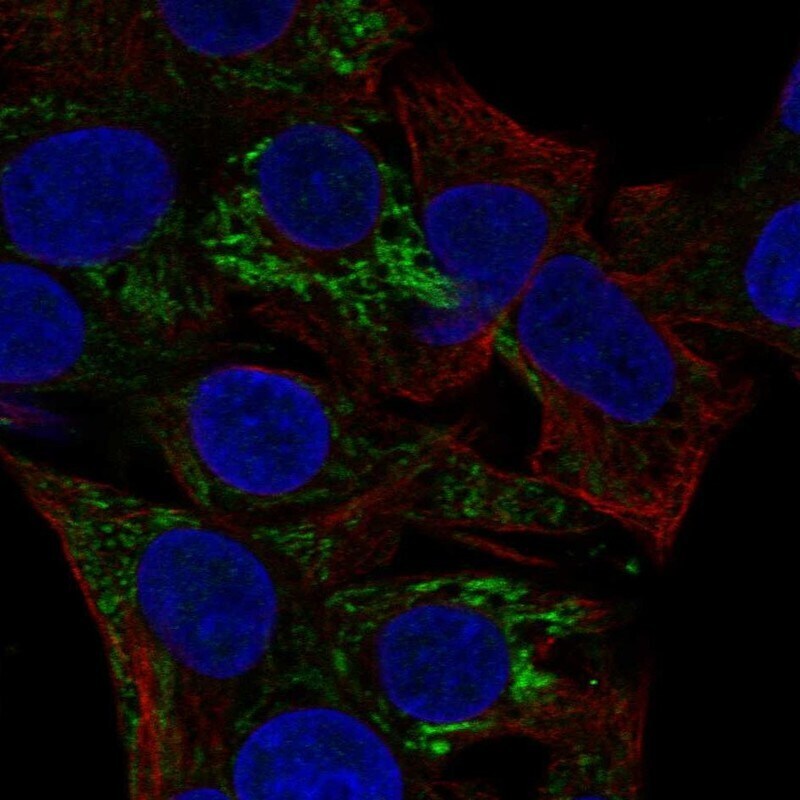
- Immunofluorescent staining of ACADS in human cell line Hep G2 using ACADS Polyclonal Antibody (Product # PA5-54580) shows localization to mitochondria.
Supportive validation
- Submitted by
- Invitrogen Antibodies (provider)
- Main image

- Experimental details
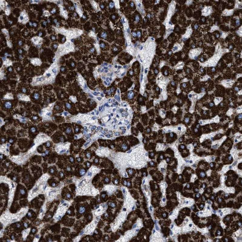
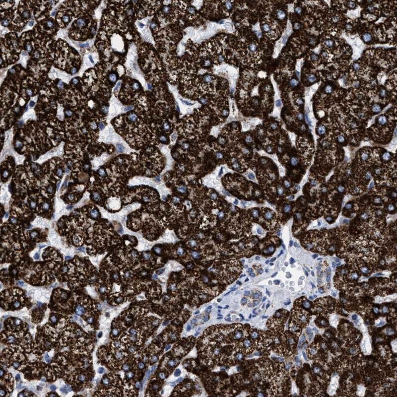
- Immunohistochemical staining of ACADS in human liver using an ACADS Polyclonal Antibody (Product # PA5-54580) shows strong cytoplasmic positivity with a granular pattern in hepatocytes and bile duct cells.
- Submitted by
- Invitrogen Antibodies (provider)
- Main image
- Experimental details
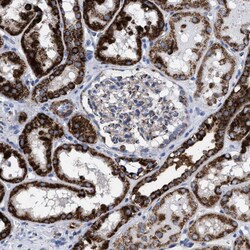
- Immunohistochemical staining of ACADS in human kidney using ACADS Polyclonal Antibody (Product # PA5-54580) shows strong cytoplasmic positivity in cells in tubules.

- Submitted by
- Invitrogen Antibodies (provider)
- Main image
- Experimental details
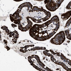
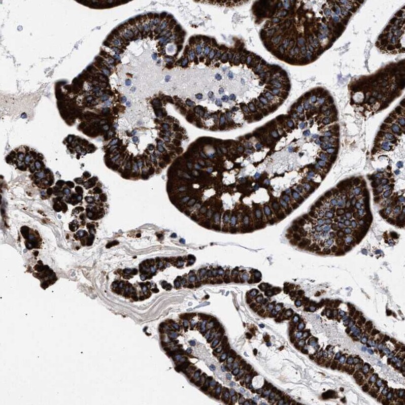
- Immunohistochemical staining of ACADS in human duodenum using ACADS Polyclonal Antibody (Product # PA5-54580) shows strong cytoplasmic positivity in glandular cells.
- Submitted by
- Invitrogen Antibodies (provider)
- Main image
- Experimental details
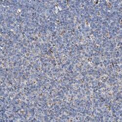
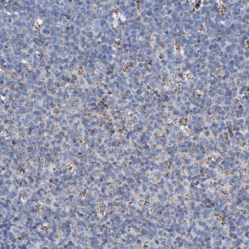
- Immunohistochemical staining of ACADS in human tonsil using ACADS Polyclonal Antibody (Product # PA5-54580) shows granular cytoplasmic positivity in germinal center cells.
- Submitted by
- Invitrogen Antibodies (provider)
- Main image
- Experimental details
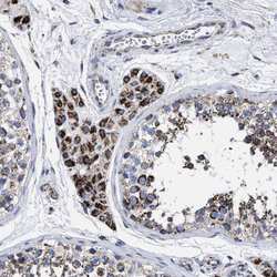
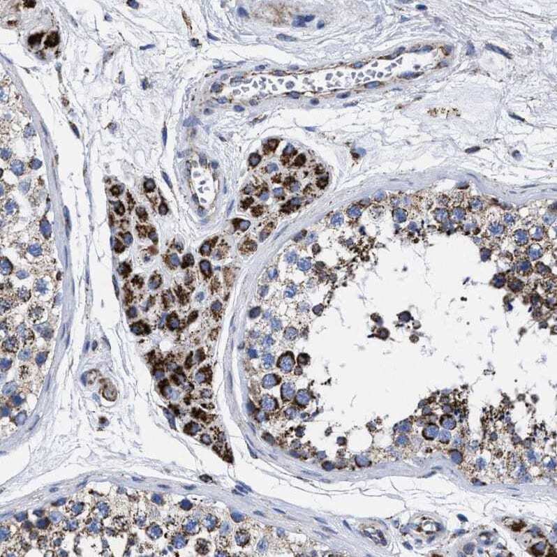
- Immunohistochemical staining of ACADS in human testis using ACADS Polyclonal Antibody (Product # PA5-54580) shows moderate cytoplasmic positivity in Leydig cells.
- Submitted by
- Invitrogen Antibodies (provider)
- Main image

- Experimental details
- Immunohistochemical staining of ACADS in human liver using ACADS Polyclonal Antibody (Product # PA5-54580) shows strong cytoplasmic positivity in hepatocytes.