Antibody data
- Antibody Data
- Antigen structure
- References [0]
- Comments [0]
- Validations
- Western blot [9]
- Immunocytochemistry [1]
Submit
Validation data
Reference
Comment
Report error
- Product number
- PA5-78544 - Provider product page
- Provider
- Invitrogen Antibodies
- Product name
- KCC2 Polyclonal Antibody
- Antibody type
- Polyclonal
- Antigen
- Synthetic peptide
- Description
- Positive Control: SK-N-SH, mouse hippocampus, rat hippocampus, rat spinal cord Predicted Reactivity: Mouse (100%) Store product as a concentrated solution. Centrifuge briefly prior to opening the vial.
- Reactivity
- Human, Mouse, Rat
- Host
- Rabbit
- Isotype
- IgG
- Vial size
- 100 µL
- Concentration
- 1.36 mg/mL
- Storage
- Store at 4°C short term. For long term storage, store at -20°C, avoiding freeze/thaw cycles.
No comments: Submit comment
Supportive validation
- Submitted by
- Invitrogen Antibodies (provider)
- Main image
- Experimental details
- Western blot analysis of KCC2 in rat tissue extract using 50 µg of protein. Samples were separated with 5% SDS-PAGE and incubated with KCC2 polyclonal antibody (Product # PA5-78544) using a dilution of 1:5000 followed by HRP-conjugated anti-rabbit IgG.
- Submitted by
- Invitrogen Antibodies (provider)
- Main image
- Experimental details
- Western blot analysis of KCC2 in mouse tissue extract using 50 µg of protein. Samples were separated with 5% SDS-PAGE and incubated with KCC2 polyclonal antibody (Product # PA5-78544) using a dilution of 1:5000 followed by HRP-conjugated anti-rabbit IgG.
- Submitted by
- Invitrogen Antibodies (provider)
- Main image
- Experimental details
- Western blot analysis of KCC2 in rat tissue extract using 50 µg of protein. Samples were separated with 5% SDS-PAGE and incubated with KCC2 polyclonal antibody (Product # PA5-78544) using a dilution of 1:5000 followed by HRP-conjugated anti-rabbit IgG.
- Submitted by
- Invitrogen Antibodies (provider)
- Main image
- Experimental details
- Western blot analysis of KCC2 in whole cell lysate using 30 µg of protein. Samples were separated with 5% SDS-PAGE and incubated with KCC2 polyclonal antibody (Product # PA5-78544) using a dilution of 1:500.
- Submitted by
- Invitrogen Antibodies (provider)
- Main image
- Experimental details
- Western Blot using KCC2 Polyclonal Antibody (Product # PA5-78544). Rat tissue extract (50 µg) was separated by 5% SDS-PAGE, and the membrane was blotted with KCC2 Polyclonal Antibody (Product # PA5-78544) diluted at 1:5,000. The HRP-conjugated anti-rabbit IgG antibody was used to detect the primary antibody.
- Submitted by
- Invitrogen Antibodies (provider)
- Main image
- Experimental details
- Western Blot using KCC2 Polyclonal Antibody (Product # PA5-78544). Mouse tissue extract (50 µg) was separated by 5% SDS-PAGE, and the membrane was blotted with KCC2 Polyclonal Antibody (Product # PA5-78544) diluted at 1:5,000. The HRP-conjugated anti-rabbit IgG antibody was used to detect the primary antibody.
- Submitted by
- Invitrogen Antibodies (provider)
- Main image
- Experimental details
- Western Blot using KCC2 Polyclonal Antibody (Product # PA5-78544). Whole cell extract (30 µg) was separated by 5% SDS-PAGE, and the membrane was blotted with KCC2 Polyclonal Antibody (Product # PA5-78544) diluted at 1:500.
- Submitted by
- Invitrogen Antibodies (provider)
- Main image
- Experimental details
- Western blot was performed using Anti-KCC2 Polyclonal Antibody (Product # PA5-78544) and ~126 kDa band corresponding to KCC2 was observed in Mouse Brain and Rat Brain, but not in Mouse Stomach and Rat Stomach which are reported to be negative. An uncharacterized band at ~45 kDa, 55 kDa were observed in the brain tissues tested. Tissue lysate (40ug lysate) of Mouse Brain (Lane 1), Rat Brain (Lane 2), Mouse Stomach (Lane 3), and Rat Stomach (Lane 4) were electrophoresed using Novex® NuPAGE® 4-12 % Bis-Tris gel (Product # NP0322BOX). Resolved proteins were then transferred onto a nitrocellulose membrane (Product # IB23001) by iBlot® 2 Dry Blotting System (Product # IB21001). The bot was probed with the primary antibody (1:1000 dilution) and detected by chemiluminescence with Goat anti-Rabbit IgG (H+L) Superclonal™ Recombinant Secondary Antibody, HRP (Product # A27036, 1:4000 dilution) using the iBright FL 1000 (Product # A32752). Chemiluminescent detection was performed using Novex® ECL Chemiluminescent Substrate Reagent Kit (Product # WP20005).
Supportive validation
- Submitted by
- Invitrogen Antibodies (provider)
- Main image
- Experimental details
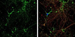
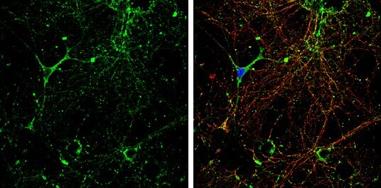
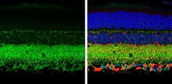
- KCC2 Polyclonal Antibody detects KCC2 protein by immunofluorescent analysis. Sample: DIV10 rat E18 primary hippocampal neurons were fixed in 4% paraformaldehyde at RT for 15 min. Green: KCC2 protein stained by KCC2 Polyclonal Antibody (Product # PA5-78544) diluted at 1:500. Red: beta Tubulin 3/ Tuj1, stained by beta-3 Tubulin Polyclonal Antibody [GT1338]diluted at 1:500. Blue: Fluoroshield with DAPI .