Explore
 Validate
 Learn
Learn Western blot
 Immunocytochemistry
Antibody data
- Antibody Data
- Antigen structure
- References [1]
- Comments [0]
- Validations
- Immunocytochemistry [2]
- Immunohistochemistry [3]
- Flow cytometry [2]
Submit
Validation data
Reference
Comment
Report error
- Product number
- PA5-72921 - Provider product page

- Provider
- Invitrogen Antibodies
- Product name
- Perilipin 1 Polyclonal Antibody
- Antibody type
- Polyclonal
- Antigen
- Synthetic peptide
- Reactivity
- Human, Mouse, Rat, Porcine
- Host
- Rabbit
- Isotype
- IgG
- Vial size
- 100 μL
- Concentration
- 1 mg/mL
- Storage
- Store at 4°C short term. For long term storage, store at -20°C, avoiding freeze/thaw cycles.
Submitted references Distinct skeletal stem cell types orchestrate long bone skeletogenesis.
Ambrosi TH, Sinha R, Steininger HM, Hoover MY, Murphy MP, Koepke LS, Wang Y, Lu WJ, Morri M, Neff NF, Weissman IL, Longaker MT, Chan CK
eLife 2021 Jul 19;10
eLife 2021 Jul 19;10
No comments: Submit comment
Supportive validation
- Submitted by
- Invitrogen Antibodies (provider)
- Main image

- Experimental details
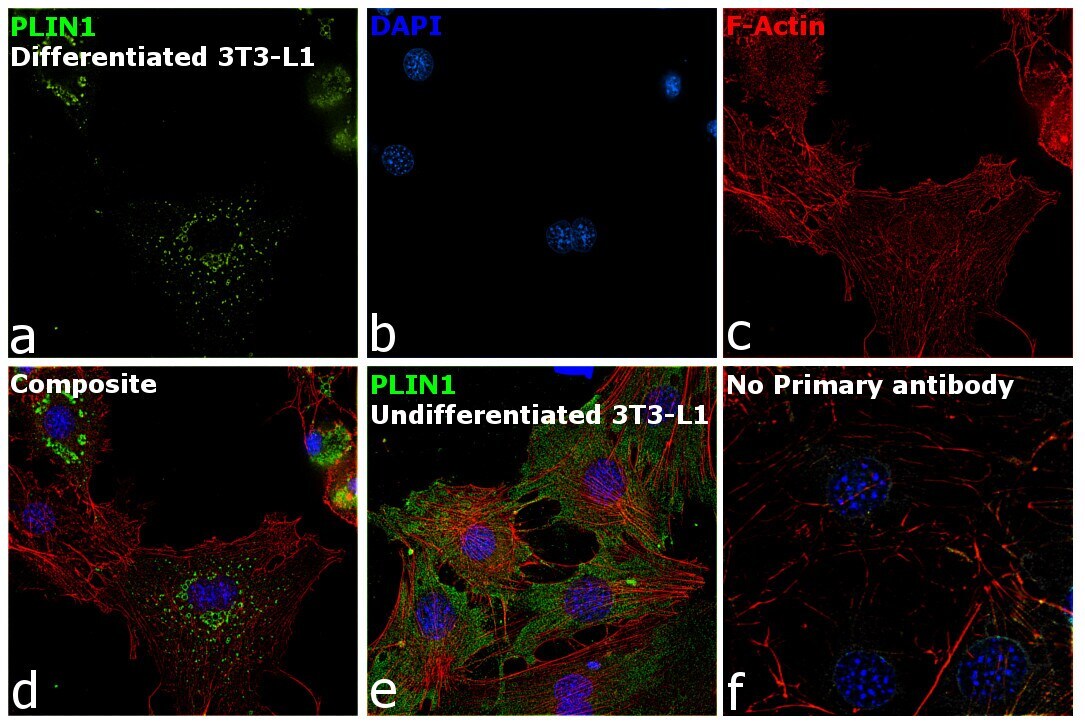
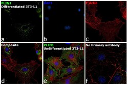
- Immunofluorescence analysis of PLIN1 was performed using 70% confluent log phase 3T3-L1 and differentiated 3T3-L1 cells. The cells were fixed with 4% paraformaldehyde for 10 minutes, permeabilized with 0.1% Triton™ X-100 for 15 minutes, and blocked with 2% BSA for 1 hour at room temperature. The cells were labeled with Perilipin 1 Polyclonal Antibody (Product # PA5-72921) at 5 µg/mL in 0.1% BSA, incubated at 4 degree Celsius overnight and then labeled with Goat anti-Rabbit IgG (H+L) Superclonal™ Recombinant Secondary Antibody, Alexa Fluor® 488 (Product # A27034) at a dilution of 1:2000 for 45 minutes at room temperature (Panel a: green). Nuclei (Panel b: blue) were stained with ProLong™ Diamond Antifade Mountant with DAPI (Product # P36962). F-actin (Panel c: red) was stained with Rhodamine Phalloidin (Product # R415). Panel d represents the merged image showing cytoplasmic localization. Panel e represents undifferentiated 3T3-L1 cells with diffused localization. Panel f represents control cells with no primary antibody to assess background The images were captured at 60X magnification.
- Submitted by
- Invitrogen Antibodies (provider)
- Main image
- Experimental details
- Immunofluorescence analysis of PLIN1 was performed using 70% confluent log phase 3T3-L1 and differentiated 3T3-L1 cells. The cells were fixed with 4% paraformaldehyde for 10 minutes, permeabilized with 0.1% Triton™ X-100 for 15 minutes, and blocked with 2% BSA for 1 hour at room temperature. The cells were labeled with Perilipin 1 Polyclonal Antibody (Product # PA5-72921) at 5 µg/mL in 0.1% BSA, incubated at 4 degree Celsius overnight and then labeled with Goat anti-Rabbit IgG (Heavy Chain) Superclonal™ Recombinant Secondary Antibody, Alexa Fluor® 488 (Product # A27034) at a dilution of 1:2000 for 45 minutes at room temperature (Panel a: green). Nuclei (Panel b: blue) were stained with ProLong™ Diamond Antifade Mountant with DAPI (Product # P36962). F-actin (Panel c: red) was stained with Rhodamine Phalloidin (Product # R415). Panel d represents the merged image showing cytoplasmic localization. Panel e represents undifferentiated 3T3-L1 cells with diffused localization. Panel f represents control cells with no primary antibody to assess background The images were captured at 60X magnification.

Supportive validation
- Submitted by
- Invitrogen Antibodies (provider)
- Main image
- Experimental details
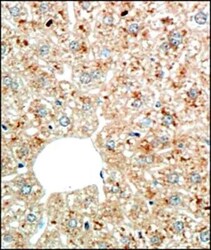
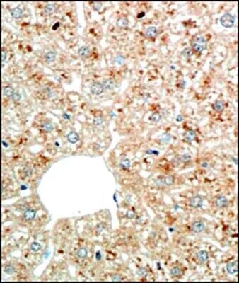
- Immunohistochemical analysis of Perilipin 1 in formalin fixed paraffin embedded section of liver tissue from mouse. Samples were incubated in Perilipin 1 polyclonal antibody (Product # PA5-72921) using a dilution of 1:200. The hepatocytes developed specific staining in the cytoplasm and around the nuclei of some cells, the immunostaining reflected a punctate appearance which is potentially the ER of the cells.
- Submitted by
- Invitrogen Antibodies (provider)
- Main image
- Experimental details
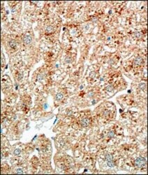
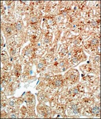
- Immunohistochemical analysis of Perilipin 1 in formalin fixed paraffin embedded section of liver tissue from mouse. Samples were incubated in Perilipin 1 polyclonal antibody (Product # PA5-72921) using a dilution of 1:200. The hepatocytes developed specific staining in the cytoplasm and around the nuclei of some cells, the immunostaining reflected a punctate appearance which is potentially the ER of the cells.
- Submitted by
- Invitrogen Antibodies (provider)
- Main image
- Experimental details
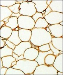
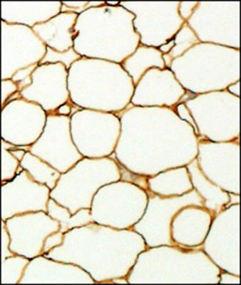
- Immunohistochemical analysis of Perilipin 1 in formalin fixed paraffin embedded section of fat tissue from mouse. Samples were incubated in Perilipin 1 polyclonal antibody (Product # PA5-72921) using a dilution of 1:200. The antibody generated an expected staining in the adipocytes towards periphery of the cells.
Supportive validation
- Submitted by
- Invitrogen Antibodies (provider)
- Main image
- Experimental details
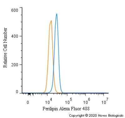
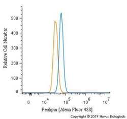
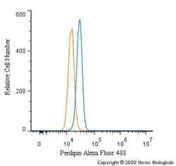
- Flow cytometry of Perilipin 1 in MCF7 cells (blue) and a matched isotype control (orange). Samples were incubated in Perilipin 1 polyclonal antibody (Product # PA5-72921) using a dilution of 10 µg/mL for 30 minutes at room temperature. Cells were fixed with 4% PFA and then permeabilized with 0.1% saponin. Both antibodies were conjugated to Alexa Fluor 488.
- Submitted by
- Invitrogen Antibodies (provider)
- Main image
- Experimental details
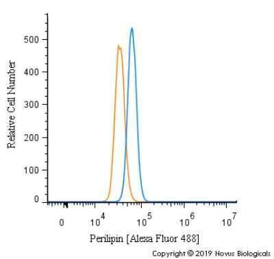
- Flow cytometry of Perilipin 1 in U2OS cells (blue) and a matched isotype control (orange). Samples were incubated in Perilipin 1 polyclonal antibody (Product # PA5-72921) using a dilution of 5 µg/mL for 30 minutes at room temperature. Cells were fixed with 4% PFA and then permeabilized with 0.1% saponin. Both antibodies were conjugated to Alexa Fluor 488.