Explore
 Validate
 Learn
Learn Western blot
 Immunocytochemistry
Antibody data
- Antibody Data
- Antigen structure
- References [1]
- Comments [0]
- Validations
- Immunocytochemistry [2]
- Immunohistochemistry [5]
Submit
Validation data
Reference
Comment
Report error
- Product number
- PA5-55046 - Provider product page

- Provider
- Invitrogen Antibodies
- Product name
- Perilipin 1 Polyclonal Antibody
- Antibody type
- Polyclonal
- Antigen
- Recombinant protein fragment
- Description
- Immunogen sequence: VCNAYEKGVQ SASSLAAWSM EPVVRRLSTQ FTAANELACR GLDHLEEKIP ALQYPPEKIA SELKDTISTR LRSARNSISV PIASTSDKVL GAALAGCELA WGVARDTAEF AANTRAGRLA SGGADLALGS IEKVVE Highest antigen sequence identity to the following orthologs: Mouse - 93%, Rat - 93%.
- Reactivity
- Human
- Host
- Rabbit
- Isotype
- IgG
- Vial size
- 100 μL
- Concentration
- 0.10 mg/mL
- Storage
- Store at 4°C short term. For long term storage, store at -20°C, avoiding freeze/thaw cycles.
Submitted references Xenogen-free isolation and culture of human adipose mesenchymal stem cells.
Doornaert M, De Maere E, Colle J, Declercq H, Taminau J, Lemeire K, Berx G, Blondeel P
Stem cell research 2019 Oct;40:101532
Stem cell research 2019 Oct;40:101532
No comments: Submit comment
Supportive validation
- Submitted by
- Invitrogen Antibodies (provider)
- Main image

- Experimental details
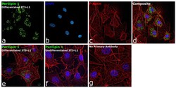
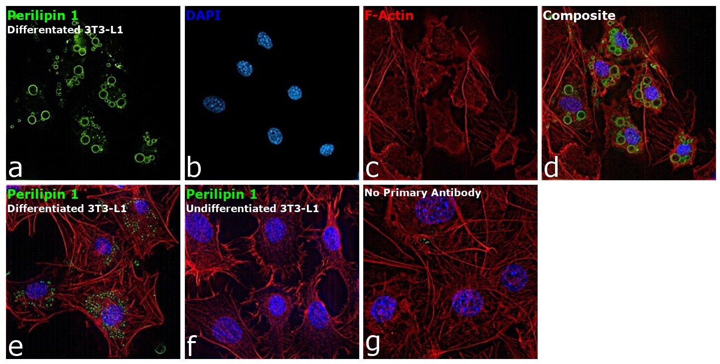
- Immunofluorescence analysis of Perilipin 1 was performed using 100% confluent log phase differentiated 3T3-L1 cells. The cells were fixed with 4% paraformaldehyde for 10 minutes, permeabilized with 0.1% Triton™ X-100 for 15 minutes, and blocked with 2% BSA for 45 minutes at room temperature. The cells were labeled with Perilipin 1 Polyclonal Antibody (Product # PA5-55046) at 1:100 dilution in 0.1% BSA, incubated at 4 degree celsius overnight and then labeled with Donkey anti-Rabbit IgG (H+L) Highly Cross-Adsorbed Secondary Antibody, Alexa Fluor Plus 488 (Product # A32790), (1:2000 dilution), for 45 minutes at room temperature (Panel a: Green). Nuclei (Panel b:Blue) were stained with ProLong™ Diamond Antifade Mountant with DAPI (Product # P36962). F-actin (Panel c: Red) was stained with Rhodamine Phalloidin (Product # R415, 1:300 dilution). Panel d and e represent the merged image showing protein localized to lipid droplets, with different stages of adipocyte differentiation. Panel f represents undifferentiated 3T3-L1 cells. Panel g represents control cells with no primary antibody to assess background. The images were captured at 60X magnification.

- Submitted by
- Invitrogen Antibodies (provider)
- Main image
- Experimental details
- Immunofluorescence analysis of Perilipin 1 was performed using 100% confluent log phase differentiated 3T3-L1 cells. The cells were fixed with 4% paraformaldehyde for 10 minutes, permeabilized with 0.1% Triton™ X-100 for 15 minutes, and blocked with 2% BSA for 45 minutes at room temperature. The cells were labeled with Perilipin 1 Polyclonal Antibody (Product # PA5-55046) at 1:100 dilution in 0.1% BSA, incubated at 4 degree celsius overnight and then labeled with Donkey anti-Rabbit IgG (H+L) Highly Cross-Adsorbed Secondary Antibody, Alexa Fluor Plus 488 (Product # A32790), (1:2000 dilution), for 45 minutes at room temperature (Panel a: Green). Nuclei (Panel b:Blue) were stained with ProLong™ Diamond Antifade Mountant with DAPI (Product # P36962). F-actin (Panel c: Red) was stained with Rhodamine Phalloidin (Product # R415, 1:300 dilution). Panel d and e represent the merged image showing protein localized to lipid droplets, with different stages of adipocyte differentiation. Panel f represents undifferentiated 3T3-L1 cells. Panel g represents control cells with no primary antibody to assess background. The images were captured at 60X magnification.
Supportive validation
- Submitted by
- Invitrogen Antibodies (provider)
- Main image

- Experimental details
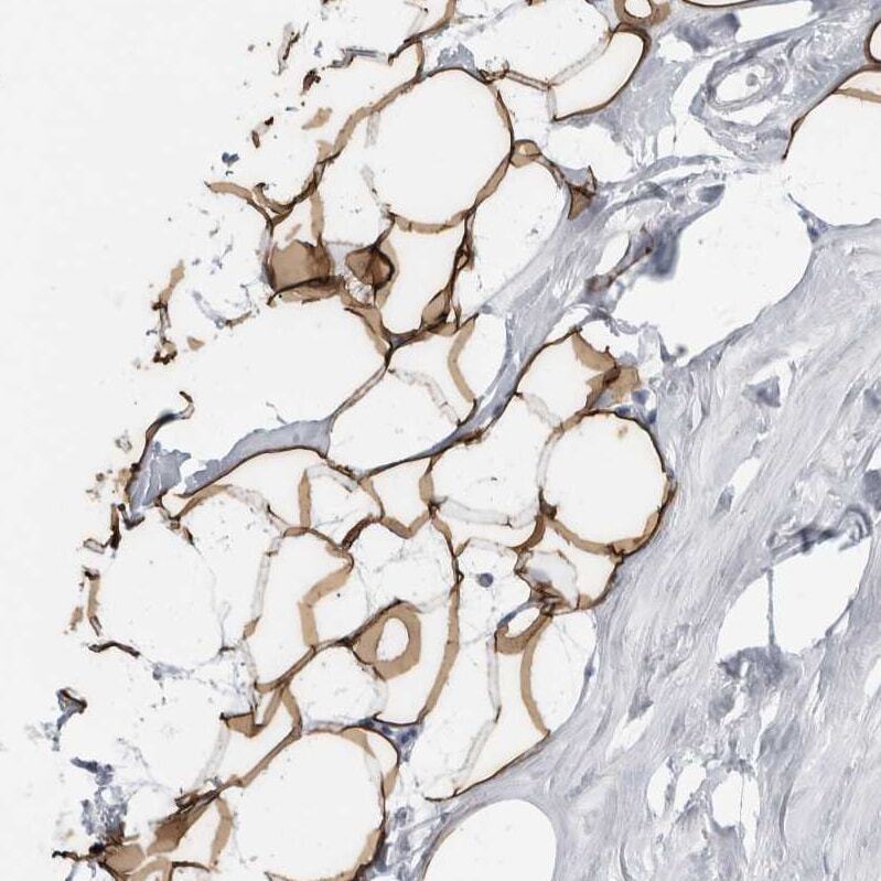
- Immunohistochemical analysis of Perilipin 1 in human adipose tissue using Perilipin 1 Polyclonal Antibody (Product # PA5-55046) shows moderate cytoplasmic positivity in adipocytes.
- Submitted by
- Invitrogen Antibodies (provider)
- Main image
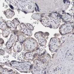
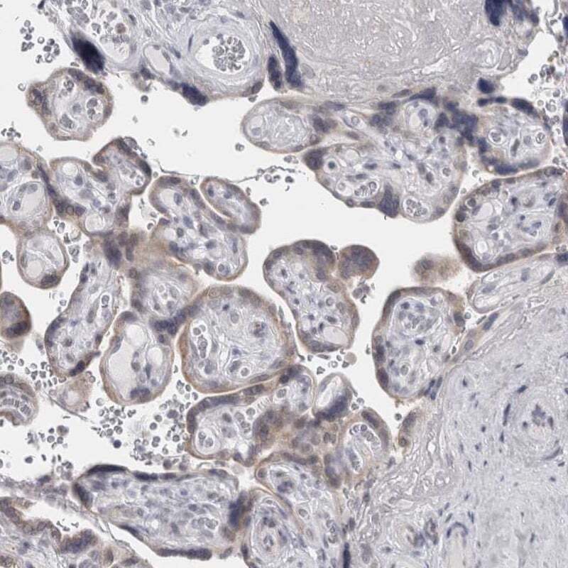
- Experimental details
- Immunohistochemical staining of Perilipin 1 in human placenta using Perilipin 1 Polyclonal Antibody (Product # PA5-55046) shows low expression as expected.
- Submitted by
- Invitrogen Antibodies (provider)
- Main image
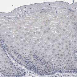
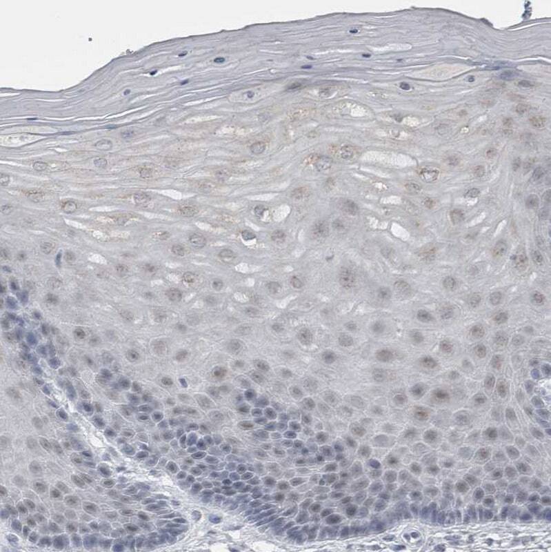
- Experimental details
- Immunohistochemical analysis of Perilipin 1 in human cervix, uterine using Perilipin 1 Polyclonal Antibody (Product # PA5-55046) shows no positivity in squamous epithelial cells as expected.
- Submitted by
- Invitrogen Antibodies (provider)
- Main image
- Experimental details
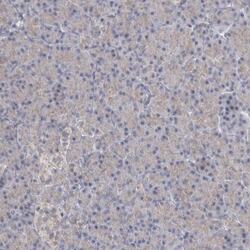
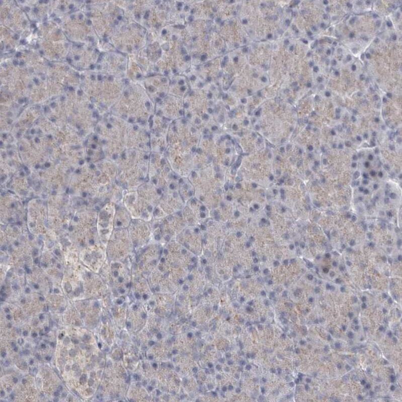
- Immunohistochemical analysis of Perilipin 1 in human pancreas using Perilipin 1 Polyclonal Antibody (Product # PA5-55046) shows very weak positivity in exocrine glandular cells.
- Submitted by
- Invitrogen Antibodies (provider)
- Main image
- Experimental details
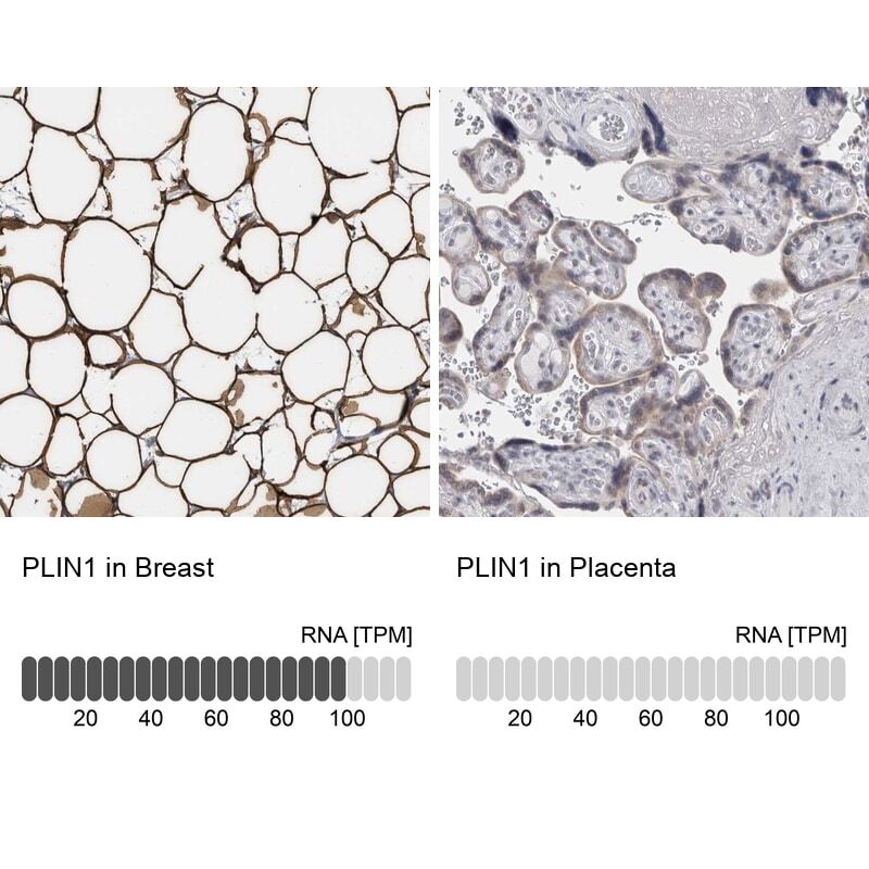
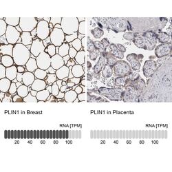
- Immunohistochemical staining of Perilipin 1 in human breast and placenta tissues using Perilipin 1 Polyclonal Antibody (Product # PA5-55046). Corresponding PLIN1 RNA-seq data are presented for the same tissues.