Explore
 Validate
 Learn
Learn Western blot
Antibody data
- Antibody Data
- Antigen structure
- References [1]
- Comments [0]
- Validations
- Western blot [2]
- Immunocytochemistry [1]
- Chromatin Immunoprecipitation [2]
Submit
Validation data
Reference
Comment
Report error
- Product number
- MA5-11187 - Provider product page

- Provider
- Invitrogen Antibodies
- Product name
- CTCF Monoclonal Antibody (G.758.4)
- Antibody type
- Monoclonal
- Antigen
- Synthetic peptide
- Description
- It is not recommended to aliquot this antibody.
- Antibody clone number
- G.758.4
- Concentration
- 91 µg/mL
Submitted references Study of mitotic chromatin supports a model of bookmarking by histone modifications and reveals nucleosome deposition patterns.
Javasky E, Shamir I, Gandhi S, Egri S, Sandler O, Rothbart SB, Kaplan N, Jaffe JD, Goren A, Simon I
Genome research 2018 Oct;28(10):1455-1466
Genome research 2018 Oct;28(10):1455-1466
No comments: Submit comment
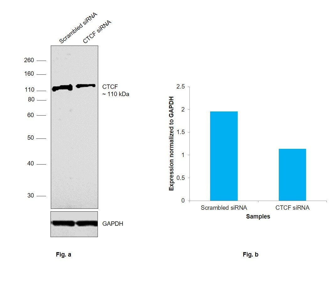
Supportive validation
- Submitted by
- Invitrogen Antibodies (provider)
- Main image

- Experimental details
- Knockdown of CTCF was achieved by transfecting MCF7 with CTCF specific siRNAs (Silencer® select Product # s20966, s20967). Western blot analysis (Fig. a) was performed using nuclear enriched extracts from the CTCF knockdown cells (lane 2) and non-targeting scrambled siRNA transfected cells (lane 1). The blot was probed with CTCF Monoclonal Antibody (G.758.4) (Product # MA5-11187, 1:1000 dilution) and Goat anti-Rabbit IgG (H+L) Superclonal™ Recombinant Secondary Antibody, HRP (Product # A27036, 1:20000 dilution). Densitometric analysis of this western blot is shown in histogram (Fig. b). Decrease in signal upon siRNA mediated knock down confirms that antibody is specific to CTCF.
- Submitted by
- Invitrogen Antibodies (provider)
- Main image

- Experimental details
- Western blot was performed using Anti-CTCF Monoclonal Antibody (G.758.4) (Product # MA5-11187) and a 110 kDa band corresponding to CTCF was observed across all the cell lines tested and as reported, decrease in the expression of CTCF was observed in MCF7 treated with Trichostatin A. An uncharacterized band (*) at ~30kDa was also found in few samples. Modified whole cell extracts (1% SDS) (30 µg lysate) of Fig (a) HeLa (Lane 1), PC-12 (Lane 2), HEK-293 (Lane 3), NIH/3T3 (Lane 4), HCT 116 (Lane 5), Jurkat (Lane 6), RAW 264.7 (Lane 7), Hep G2 (Lane 8); Fig (b) MCF7 (Lane 1) and MCF7 treated with Trichostatin A (10uM for 24hr) (Lane 2) were electrophoresed using Novex® NuPAGE® 4-12% Bis-Tris Protein Gel (Product # NP0321BOX). Resolved proteins were then transferred onto a nitrocellulose membrane (Product # IB23001) by iBlot® 2 Dry Blotting System (Product # IB21001). The blot was probed with the primary antibody (1:1000 dilution) and detected by chemiluminescence with Goat anti-Rabbit IgG (H+L), Superclonal™ Recombinant Secondary Antibody, HRP (Product # A27036, 1:4000 dilution) using the iBright FL 1000 (Product # A32752). Chemiluminescent detection was performed using Novex® ECL Chemiluminescent Substrate Reagent Kit (Product # WP20005).

Supportive validation
- Submitted by
- Invitrogen Antibodies (provider)
- Main image

- Experimental details
- Immunofluorescence analysis of CTCF was performed using 70% confluent log phase MCF7 cells. The cells were fixed with 4% paraformaldehyde for 10 minutes, permeabilized with 0.1% Triton™ X-100 for 15 minutes, and blocked with 2% BSA for 1 hour at room temperature. The cells were labeled with CTCF Monoclonal Antibody (G.758.4) (Product # MA5-11187) at 1:100 dilution in 0.1% BSA, incubated at 4 degree celsius overnight and then labeled with Donkey anti-Rabbit IgG (H+L) Highly Cross-Adsorbed Secondary Antibody, Alexa Fluor Plus 488 (Product # A32790), (1:2000 dilution), for 45 minutes at room temperature (Panel a: Green). Nuclei (Panel b:Blue) were stained with ProLong™ Diamond Antifade Mountant with DAPI (Product # P36962). F-actin (Panel c: Red) was stained with Rhodamine Phalloidin (Product # R415, 1:300). Panel d represents the merged image showing nuclear localization. Panel e represents control cells with no primary antibody to assess background. The images were captured at 60X magnification.

Supportive validation
- Submitted by
- Invitrogen Antibodies (provider)
- Main image

- Experimental details
- Chromatin Immunoprecipitation (ChIP) of CTCF protein was performed using CTCF Monoclonal Antibody (G.758.4) (Product # MA5-11187, 10 µL) on sheared chromatin from MCF7 cells using the MAGnify ChIP System kit (Product # 49-2024). Normal Rabbit IgG was used as a negative IP control. The purified DNA was analyzed by qPCR using primers binding to H19-ICR, CRY1, PFKFB3, GLI2 (5), AXL (3) (Active), EXON4/5 OF H19 and SAT2 satellite repeats (Inactive). Data is presented as fold enrichment of the antibody signal versus the negative control IgG using the comparative CT method.

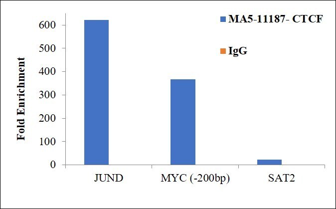
- Submitted by
- Invitrogen Antibodies (provider)
- Main image

- Experimental details
- Chromatin Immunoprecipitation (ChIP) assay of endogenous CTCF protein using Anti-CTCF Antibody: ChIP was performed using Anti-CTCF Monoclonal Antibody (G.758.4) (Product # MA5-11187) 5 µg, on sheared chromatin from HCT116 cells using the MAGnify ChIP System kit (Product # 49-2024). Normal Rabbit IgG was used as a negative IP control. The purified DNA was analyzed by qPCR using primers binding to promoter of MYC, JUND and SAT2 satellite repeats. Data is presented as fold enrichment of the antibody signal versus the negative control IgG using the comparative CT method.