Explore
 Validate
 Learn
Learn Western blot
 Immunoprecipitation
Antibody data
- Antibody Data
- Antigen structure
- References [0]
- Comments [0]
- Validations
- Western blot [4]
- Immunocytochemistry [4]
- Immunohistochemistry [6]
- Flow cytometry [1]
Submit
Validation data
Reference
Comment
Report error
- Product number
- MA5-32640 - Provider product page

- Provider
- Invitrogen Antibodies
- Product name
- CTCF Recombinant Rabbit Monoclonal Antibody (JM10-61)
- Antibody type
- Monoclonal
- Antigen
- Synthetic peptide
- Description
- Recombinant rabbit monoclonal antibodies are produced using in vitro expression systems. The expression systems are developed by cloning in the specific antibody DNA sequences from immunoreactive rabbits. Then, individual clones are screened to select the best candidates for production. The advantages of using recombinant rabbit monoclonal antibodies include: better specificity and sensitivity, lot-to-lot consistency, animal origin-free formulations, and broader immunoreactivity to diverse targets due to larger rabbit immune repertoire.
- Reactivity
- Human, Mouse, Rat, Zebrafish
- Host
- Rabbit
- Isotype
- IgG
- Antibody clone number
- JM10-61
- Vial size
- 100 µL
- Concentration
- 1 mg/mL
- Storage
- Store at 4°C short term. For long term storage, store at -20°C, avoiding freeze/thaw cycles.
No comments: Submit comment
Supportive validation
- Submitted by
- Invitrogen Antibodies (provider)
- Main image

- Experimental details
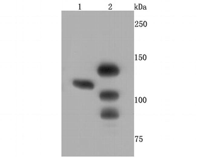
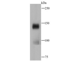
- Western blot analysis of CTCF in different lysates using a Monoclonal antibody (Product #MA5-32640) at a dilution of 1:500. Positive control: Lane 1: 293T, Lane 2:MCF-7.
- Submitted by
- Invitrogen Antibodies (provider)
- Main image
- Experimental details
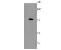
- Western blot analysis of CTCF in Zebrafish tissue lysates using a CTCF Monoclonal antibody (Product # MA5-32640) at a dilution of 1:200.
- Submitted by
- Invitrogen Antibodies (provider)
- Main image
- Experimental details
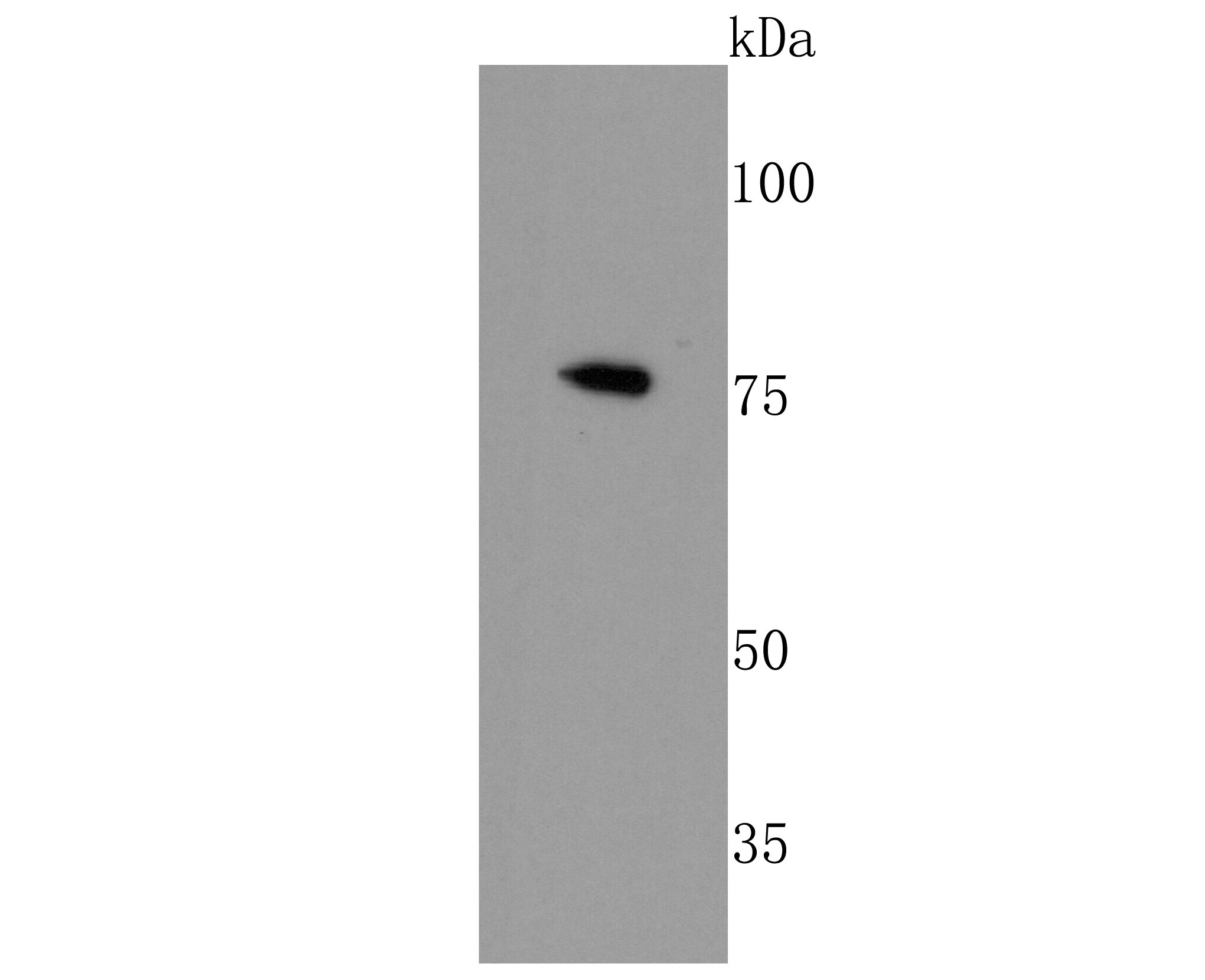
- Western blot analysis of CTCF in hybrid fish (crucian-carp) brain tissue lysate using a CTCF Monoclonal antibody (Product # MA5-32640) at a dilution of 1:500.

- Submitted by
- Invitrogen Antibodies (provider)
- Main image
- Experimental details
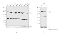
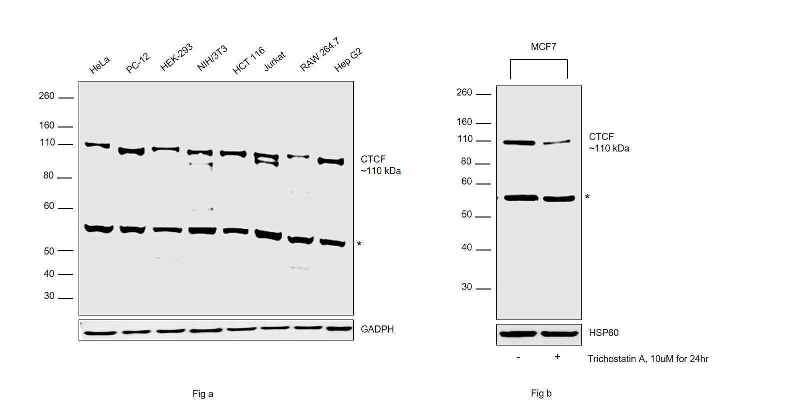
- Western blot was performed using Anti-CTCF Recombinant Rabbit Monoclonal Antibody (JM10-61) (Product # MA5-32640) and a 110 kDa band corresponding to CTCF and an uncharacterized band (*) at ~55kDa was observed across all the cell lines tested. Modified whole cell extracts (1% SDS) (30 µg lysate) of Fig (a) HeLa (Lane 1), PC-12 (Lane 2), HEK-293 (Lane 3), NIH/3T3 (Lane 4), HCT 116 (Lane 5), Jurkat (Lane 6), RAW 264.7 (Lane 7), Hep G2 (Lane 8); Fig (b) MCF7 (Lane 1) and MCF7 treated with Trichostatin A (10uM for 24hr) (Lane 2) were electrophoresed using Novex® NuPAGE® 4-12% Bis-Tris Protein Gel (Product # NP0321BOX). Resolved proteins were then transferred onto a nitrocellulose membrane (Product # IB23001) by iBlot® 2 Dry Blotting System (Product # IB21001). The blot was probed with the primary antibody (1:500 dilution) and detected by chemiluminescence with Goat anti-Rabbit IgG (H+L), Superclonal™ Recombinant Secondary Antibody, HRP (Product # A27036, 1:4000 dilution) using the iBright FL 1000 (Product # A32752). Chemiluminescent detection was performed using Novex® ECL Chemiluminescent Substrate Reagent Kit (Product # WP20005).
Supportive validation
- Submitted by
- Invitrogen Antibodies (provider)
- Main image
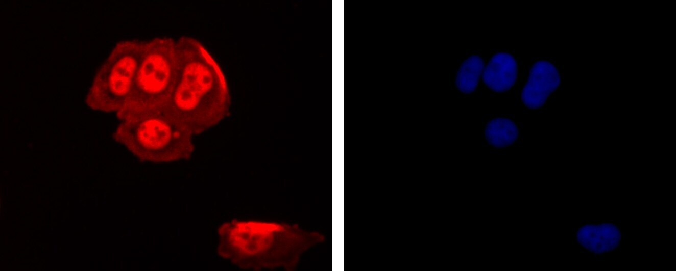
- Experimental details
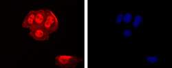
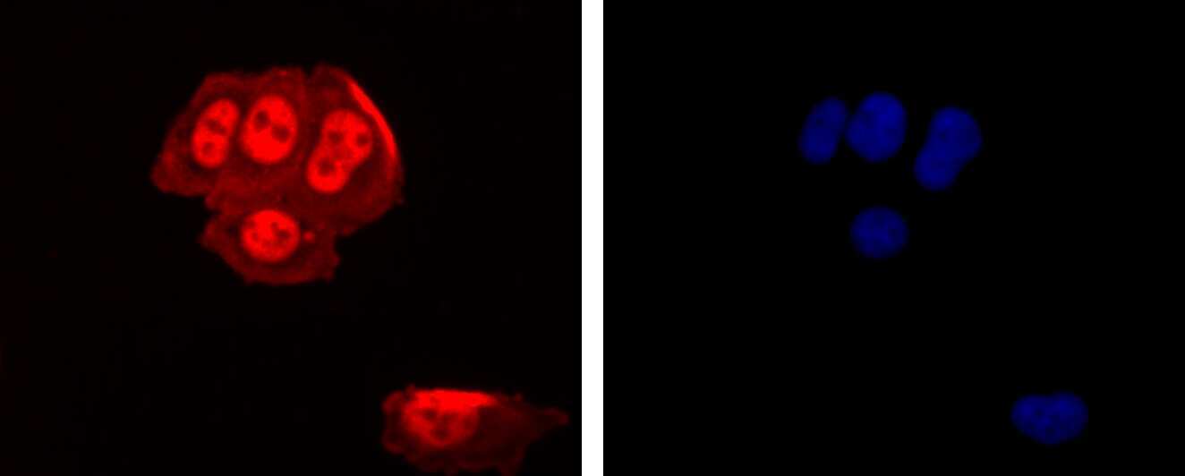
- Immunocytochemical analysis of CTCF in MCF-7 cells using a CTCF Monoclonal antibody (Product # MA5-32640) as seen in red. The nuclear counter stain is DAPI (blue). Cells were fixed in paraformaldehyde, permeabilised with 0.25% Triton X100/PBS.
- Submitted by
- Invitrogen Antibodies (provider)
- Main image
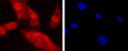
- Experimental details
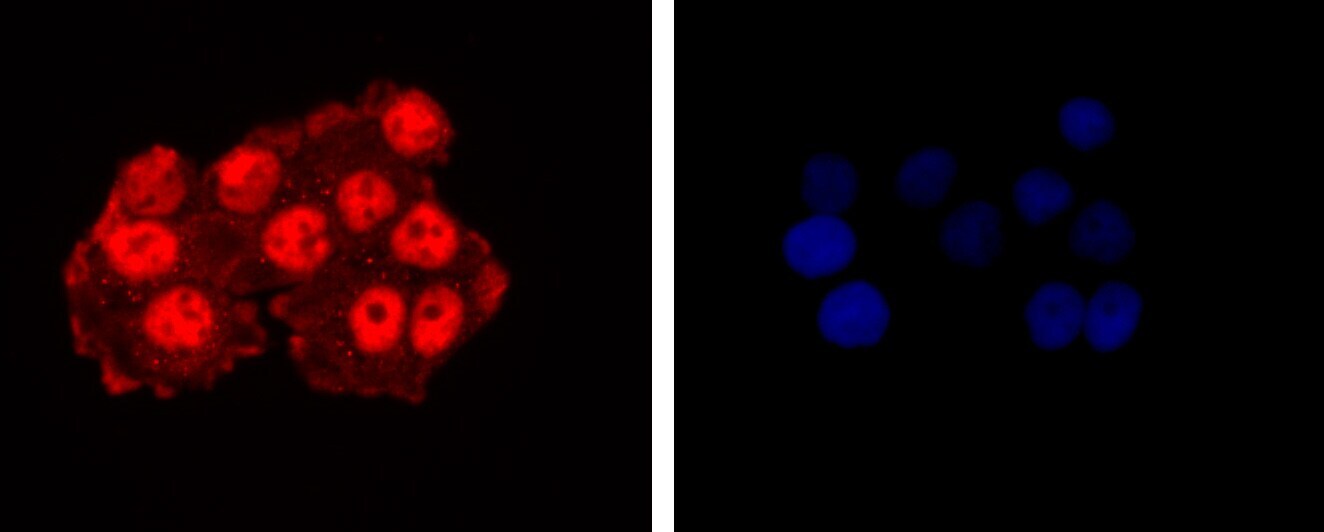
- Immunocytochemical analysis of CTCF in Hela cells using a CTCF Monoclonal antibody (Product # MA5-32640) as seen in red. The nuclear counter stain is DAPI (blue). Cells were fixed in paraformaldehyde, permeabilised with 0.25% Triton X100/PBS.
- Submitted by
- Invitrogen Antibodies (provider)
- Main image
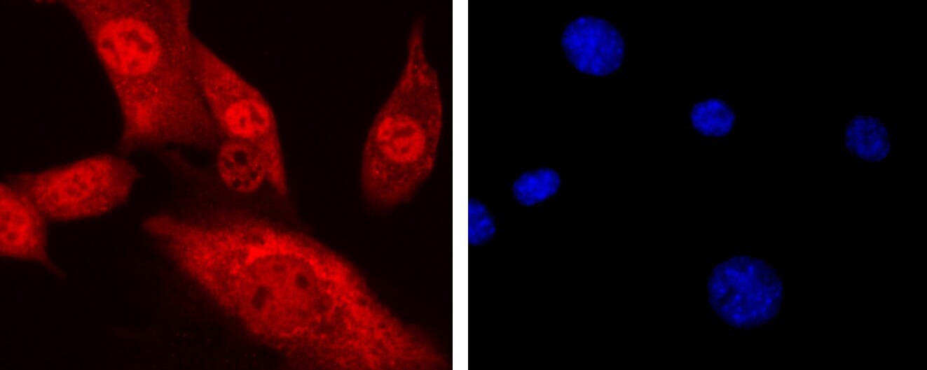
- Experimental details
- Immunocytochemical analysis of CTCF in MCF-7 cells using a CTCF Monoclonal antibody (Product # MA5-32640) as seen in red. The nuclear counter stain is DAPI (blue). Cells were fixed in paraformaldehyde, permeabilised with 0.25% Triton X100/PBS.
- Submitted by
- Invitrogen Antibodies (provider)
- Main image
- Experimental details
- Immunocytochemical analysis of CTCF in NIH-3T3 cells using a CTCF Monoclonal antibody (Product # MA5-32640) as seen in red. The nuclear counter stain is DAPI (blue). Cells were fixed in paraformaldehyde, permeabilised with 0.25% Triton X100/PBS.
Supportive validation
- Submitted by
- Invitrogen Antibodies (provider)
- Main image
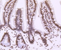
- Experimental details
- Immunohistochemical analysis of CTCF of paraffin-embedded Mouse colon tissue using a CTCF Monoclonal antibody (Product #MA5-32640). Counter stained with hematoxylin.

- Submitted by
- Invitrogen Antibodies (provider)
- Main image
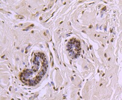
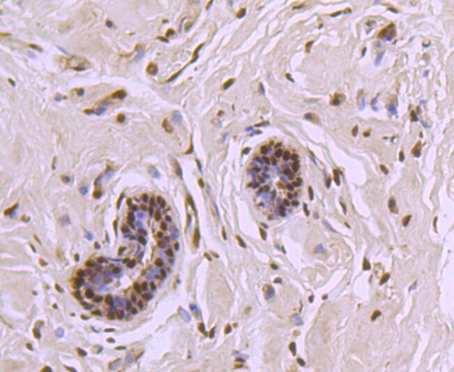
- Experimental details
- Immunohistochemical analysis of CTCF of paraffin-embedded Human breast carcinoma tissue using a CTCF Monoclonal antibody (Product #MA5-32640). Counter stained with hematoxylin.
- Submitted by
- Invitrogen Antibodies (provider)
- Main image

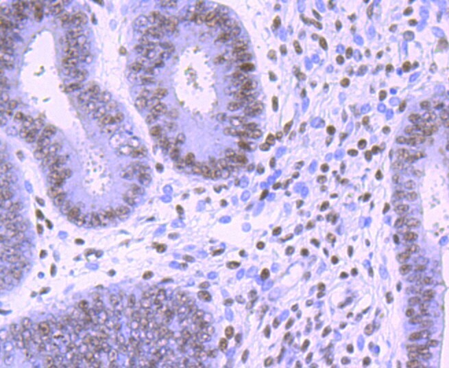
- Experimental details
- Immunohistochemical analysis of CTCF of paraffin-embedded Human endometrium tissue using a CTCF Monoclonal antibody (Product #MA5-32640). Counter stained with hematoxylin.
- Submitted by
- Invitrogen Antibodies (provider)
- Main image
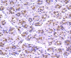
- Experimental details
- Immunohistochemical analysis of CTCF of paraffin-embedded Mouse stomach tissue using a CTCF Monoclonal antibody (Product #MA5-32640). Counter stained with hematoxylin.

- Submitted by
- Invitrogen Antibodies (provider)
- Main image

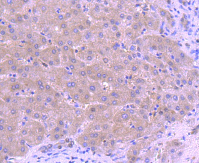
- Experimental details
- Immunohistochemical analysis of CTCF of paraffin-embedded Human liver tissue using a CTCF Monoclonal antibody (Product #MA5-32640). Counter stained with hematoxylin.
- Submitted by
- Invitrogen Antibodies (provider)
- Main image
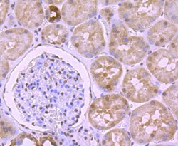
- Experimental details
- Immunohistochemical analysis of CTCF of paraffin-embedded Human kidney tissue using a CTCF Monoclonal antibody (Product #MA5-32640). Counter stained with hematoxylin.

Supportive validation
- Submitted by
- Invitrogen Antibodies (provider)
- Main image
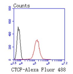
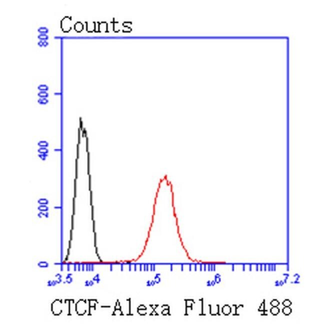
- Experimental details
- Flow Cytometric analysis of CTCF in 293T cells using a CTCF Monoclonal Antibody (Product # MA5-32640) at a dilution of 1:50, as seen in red compared with an unlabelled control (cells without incubation with primary antibody; black). Alexa Fluor 488-conjugated goat anti rabbit IgG was used as the secondary antibody..