Explore
 Validate
 Learn
Learn Western blot
Antibody data
- Antibody Data
- Antigen structure
- References [0]
- Comments [0]
- Validations
- Western blot [4]
- Immunocytochemistry [2]
- Immunohistochemistry [1]
Submit
Validation data
Reference
Comment
Report error
- Product number
- PA5-54274 - Provider product page

- Provider
- Invitrogen Antibodies
- Product name
- DPF2 Polyclonal Antibody
- Antibody type
- Polyclonal
- Antigen
- Recombinant full-length protein
- Description
- Immunogen sequence: SFPSIKPDTD QTLKKEGLIS QDGSSLEALL RTDPLEKRGA PDPRVDDDSL GEFPVTNSRA RKRILEPDDF LDDLDDEDYE EDTPKRRGKG KSKGKGVGS Highest antigen sequence identity to the following orthologs: Mouse - 96%, Rat - 98%.
- Reactivity
- Human
- Host
- Rabbit
- Isotype
- IgG
- Vial size
- 100 µL
- Concentration
- 0.05 mg/mL
- Storage
- Store at 4°C short term. For long term storage, store at -20°C, avoiding freeze/thaw cycles.
No comments: Submit comment
Supportive validation
- Submitted by
- Invitrogen Antibodies (provider)
- Main image
- Experimental details
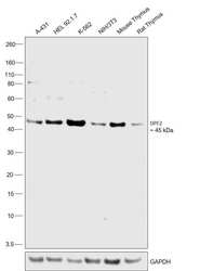
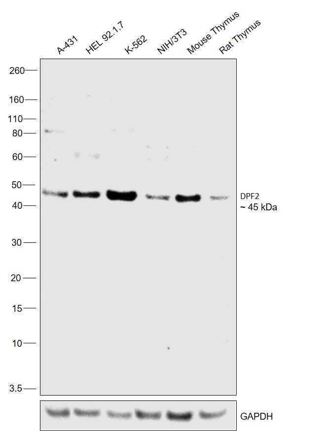
- Western blot was performed using Anti-DPF2 Polyclonal Antibody (Product # PA5-54274) and a 45 kDa band corresponding to Zinc finger protein ubi-d4 was observed across cell lines and tissues tested. Nuclear enriched extracts (30 µg lysate) of A-431 (Lane 1), HEL 92.1.7 (Lane 2), K-562 (Lane 3), NIH/3T3 (Lane 4). Tissue extracts (30 µg lysate) of Mouse Thymus (Lane 5) and Rat Thymus (Lane 6) were electrophoresed using NuPAGE™ 4-12% Bis-Tris Protein Gel (Product # NP0322BOX). Resolved proteins were then transferred onto a nitrocellulose membrane (Product # IB23001) by iBlot® 2 Dry Blotting System (Product # IB21001). The blot was probed with the primary antibody (0.4 µg/mL) and detected by chemiluminescence with Goat anti-Rabbit IgG (H+L) Superclonal™ Recombinant Secondary Antibody, HRP (Product # A27036,1:4000 dilution) using the iBright FL 1000 (Product # A32752). Chemiluminescent detection was performed using Novex® ECL Chemiluminescent Substrate Reagent Kit (Product # WP20005).
- Submitted by
- Invitrogen Antibodies (provider)
- Main image
- Experimental details
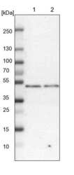
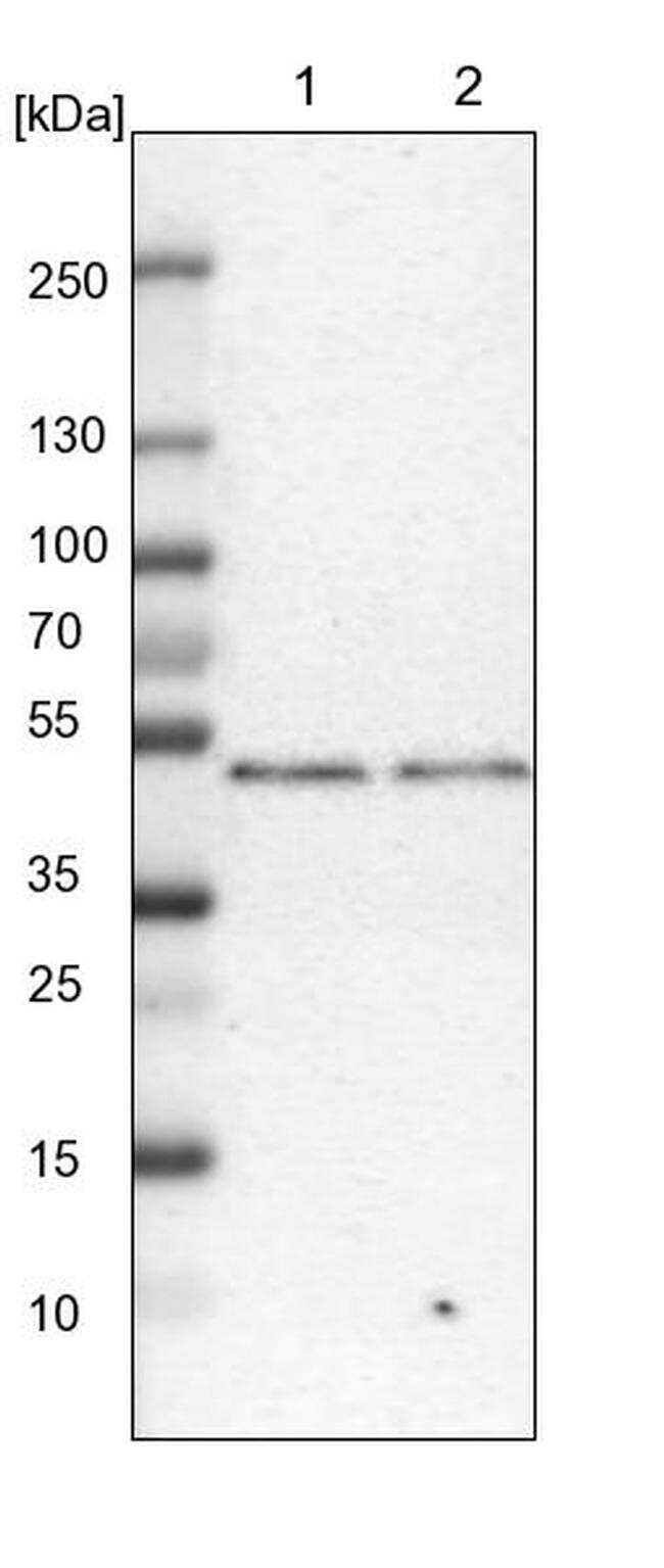
- Western blot analysis of DPF2 in Lane 1: NIH-3T3 cell lysate (Mouse embryonic fibroblast cells); Lane 2: NBT-II cell lysate (Rat Wistar bladder tumour cells). Samples were probed using a DPF2 Polyclonal Antibody (Product # PA5-54274).
- Submitted by
- Invitrogen Antibodies (provider)
- Main image

- Experimental details
- Western blot was performed using Anti-DPF2 Polyclonal Antibody (Product # PA5-54274) and a 45 kDa band corresponding to Zinc finger protein ubi-d4 was observed across cell lines and tissues tested. Nuclear enriched extracts (30 µg lysate) of A-431 (Lane 1), HEL 92.1.7 (Lane 2), K-562 (Lane 3), NIH/3T3 (Lane 4). Tissue extracts (30 µg lysate) of Mouse Thymus (Lane 5) and Rat Thymus (Lane 6) were electrophoresed using NuPAGE™ 4-12% Bis-Tris Protein Gel (Product # NP0322BOX). Resolved proteins were then transferred onto a nitrocellulose membrane (Product # IB23001) by iBlot® 2 Dry Blotting System (Product # IB21001). The blot was probed with the primary antibody (0.4 µg/mL) and detected by chemiluminescence with Goat anti-Rabbit IgG (H+L) Superclonal™ Recombinant Secondary Antibody, HRP (Product # A27036,1:4000 dilution) using the iBright FL 1000 (Product # A32752). Chemiluminescent detection was performed using Novex® ECL Chemiluminescent Substrate Reagent Kit (Product # WP20005).

- Submitted by
- Invitrogen Antibodies (provider)
- Main image

- Experimental details
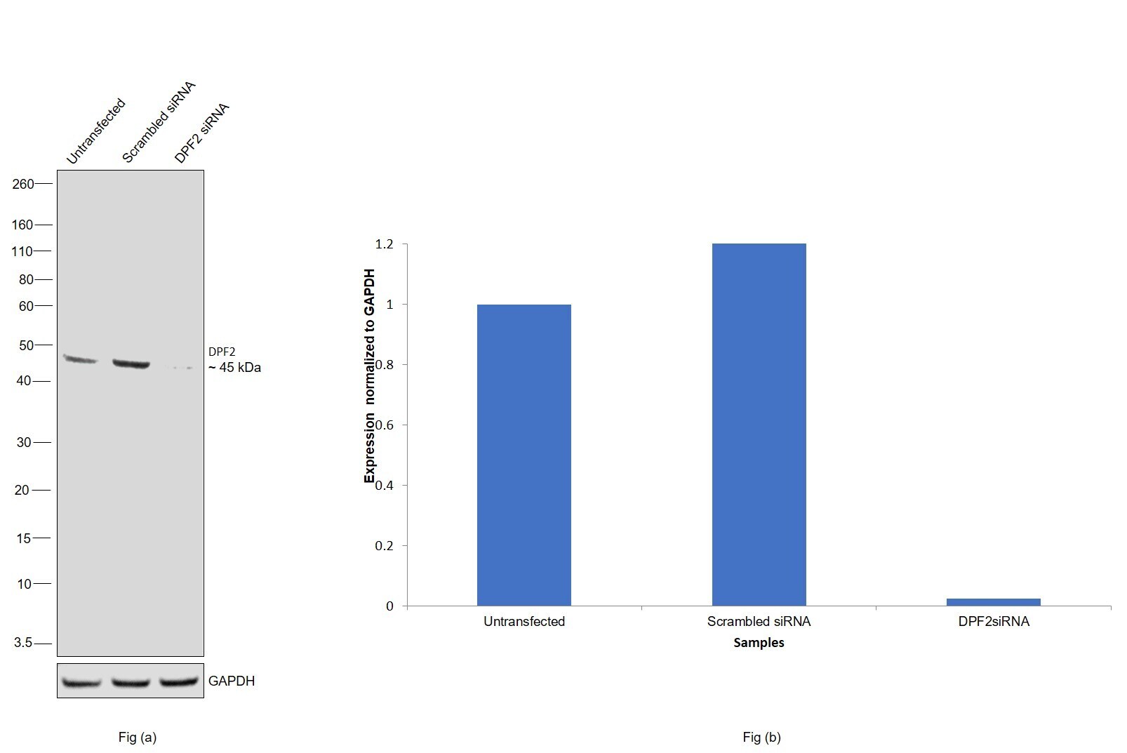
- Knockdown of Zinc finger protein ubi-d4 was achieved by transfecting K-562 with Zinc finger protein ubi-d4 specific siRNAs (Silencer® select Product # S11929, S11930). Western blot analysis (Fig. a) was performed using Nuclear enriched extracts from the Zinc finger protein ubi-d4 knockdown cells (lane 3), non-targeting scrambled siRNA transfected cells (lane 2) and untransfected cells (lane 1). The blot was probed with DPF2 Polyclonal Antibody (Product # PA5-54274, 0.4 µg/mL ) and Goat anti-Rabbit IgG (H+L) Superclonal™ Recombinant Secondary Antibody, HRP (Product # A27036, 1:4000 dilution). Densitometric analysis of this western blot is shown in histogram (Fig. b). Decrease in signal upon siRNA mediated knock down confirms that antibody is specific to Zinc finger protein ubi-d4.
Supportive validation
- Submitted by
- Invitrogen Antibodies (provider)
- Main image
- Experimental details
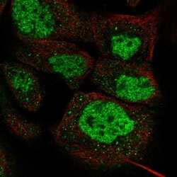
- Immunofluorescent staining of DPF2 in human cell line A-431 shows positivity in nucleus but excluded from the nucleoli. Samples were probed using a DPF2 Polyclonal Antibody (Product # PA5-54274).

- Submitted by
- Invitrogen Antibodies (provider)
- Main image
- Experimental details
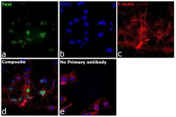
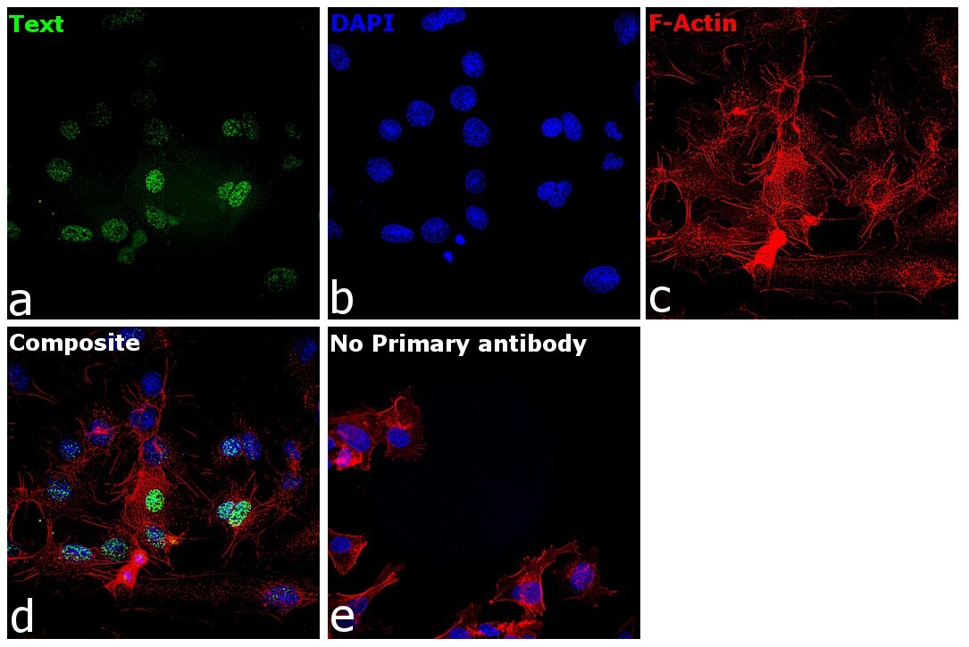
- Immunofluorescence analysis of Zinc finger protein ubi-d4 was performed using 70% confluent log phase Hep G2 cells. The cells were fixed with 4% paraformaldehyde for 10 minutes, permeabilized with 0.1% Triton™ X-100 for 15 minutes, and blocked with 2% BSA for 45 minutes at room temperature. The cells were labeled with DPF2 Polyclonal Antibody (Product # PA5-54274) at 1:100 dilution in 0.1% BSA, incubated at 4 degree celsius overnight and then labeled with Donkey anti-Rabbit IgG (H+L) Highly Cross-Adsorbed Secondary Antibody, Alexa Fluor Plus 488 (Product # A32790), (1:2000 dilution), for 45 minutes at room temperature (Panel a: Green). Nuclei (Panel b:Blue) were stained with Hoechst 33342 (Product # H1399). F-actin (Panel c: Red) was stained with Rhodamine Phalloidin (Product # R415, 1:300 dilution). Panel d represents the merged image showing Nuclear localization. Panel e represents control cells with no primary antibody to assess background. The images were captured at 40X magnification in CellInsight CX7 LZR High-Content Screening (HCS) Platform (Product # CX7A1110LZR) and externally deconvoluted (D.Sage et al. / Methods 115 (2017) 28–41).
Supportive validation
- Submitted by
- Invitrogen Antibodies (provider)
- Main image

- Experimental details
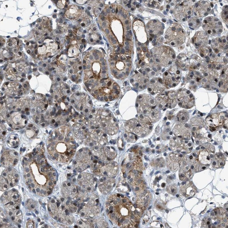
- Immunohistochemical staining of DPF2 in human salivary gland using a DPF2 Polyclonal Antibody (Product # PA5-54274) shows weak cytoplasmic and nuclear positivity in glandular cells.