 Explore
 Validate
 Learn
Learn Western blot
Antibody data
- Antibody Data
- Antigen structure
- References [0]
- Comments [0]
- Validations
- Western blot [6]
- Immunocytochemistry [4]
- Immunohistochemistry [2]
Submit
Validation data
Reference
Comment
Report error
- Product number
- MA5-17271 - Provider product page

- Provider
- Invitrogen Antibodies
- Product name
- IDH2 Monoclonal Antibody (GT673)
- Antibody type
- Monoclonal
- Antigen
- Recombinant protein fragment
- Description
- Recommended positive controls: HepG2, Jurkat, Raji, K562, THP-1, NCI-H929, mouse brain, rat brain, DDDDK-tagged IDH2-transfected 293T. Predicted reactivity: Mouse (97%), Rat (96%), Zebrafish (89%), Xenopus laevis (91%), Pig (96%), Chicken (95%), Rhesus Monkey (100%), Bovine (96%). Store product as a concentrated solution. Centrifuge briefly prior to opening the vial.
- Reactivity
- Human, Mouse, Rat
- Host
- Mouse
- Isotype
- IgG
- Antibody clone number
- GT673
- Vial size
- 100 µL
- Concentration
- 1 mg/mL
- Storage
- Store at 4°C short term. For long term storage, store at -20°C, avoiding freeze/thaw cycles.
No comments: Submit comment
Supportive validation
- Submitted by
- Invitrogen Antibodies (provider)
- Main image

- Experimental details
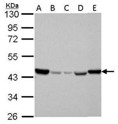
- IDH2 Polyclonal Antibody detects IDH2 protein by western blot analysis. A. 30 µg Jurkat whole cell lysate/extract. B. 30 µg Raji whole cell lysate/extract. C. 30 µg K562 whole cell lysate/extract. D. 30 µg THP-1 whole cell lysate/extract. E. 30 µg NCI-H929 whole cell lysate/extract.10% SDS-PAGE. IDH2 Polyclonal Antibody IDH2 Monoclonal Antibody (GT673) (Product # MA5-17271) dilution: 1:1,000. The HRP-conjugated anti-mouse IgG antibody was used to detect the primary antibody.

- Submitted by
- Invitrogen Antibodies (provider)
- Main image
- Experimental details
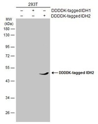
- Western Blot analysis of IDH2 was performed by separating 30 µg of non-transfected (–) and transfected (+) 293T whole cell extracts by 5% SDS-PAGE. Proteins were transferred to a membrane and probed with a IDH2 Monoclonal Antibody (GT673) (Product # MA5-17271) at a dilution of 1:1000. The HRP-conjugated anti-mouse IgG antibody was used to detect the primary antibody.

- Submitted by
- Invitrogen Antibodies (provider)
- Main image
- Experimental details
- IDH2 Polyclonal Antibody detects IDH2 protein by western blot analysis. A. 30 µg Jurkat whole cell lysate/extract. B. 30 µg Raji whole cell lysate/extract. C. 30 µg K562 whole cell lysate/extract. D. 30 µg THP-1 whole cell lysate/extract. E. 30 µg NCI-H929 whole cell lysate/extract.10% SDS-PAGE. IDH2 Polyclonal Antibody IDH2 Monoclonal Antibody (GT673) (Product # MA5-17271) dilution: 1:1,000. The HRP-conjugated anti-mouse IgG antibody was used to detect the primary antibody.

- Submitted by
- Invitrogen Antibodies (provider)
- Main image
- Experimental details
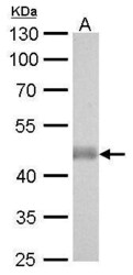
- IDH2 Monoclonal Antibody (GT673) detects IDH2 protein by western blot analysis. A. 50 µg mouse brain lysate/extract.10% SDS-PAGE. IDH2 Monoclonal Antibody (GT673) (Product # MA5-17271) dilution: 1:1,000. The HRP-conjugated anti-mouse IgG antibody was used to detect the primary antibody.

- Submitted by
- Invitrogen Antibodies (provider)
- Main image
- Experimental details
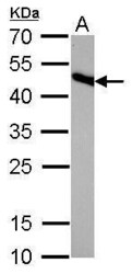
- IDH2 Monoclonal Antibody (GT673) detects IDH2 protein by western blot analysis. A. 50 µg rat brain lysate/extract.12% SDS-PAGE. IDH2 Monoclonal Antibody (GT673) (Product # MA5-17271) dilution: 1:1,000. The HRP-conjugated anti-mouse IgG antibody was used to detect the primary antibody.

- Submitted by
- Invitrogen Antibodies (provider)
- Main image

- Experimental details
- Western blot analysis was performed on whole cell extracts (30 µg lysate) of Hep G2 (Lane 1), MCF7 (Lane 2), U-87 MG (Lane 3), LNCaP (Lane 4), C2C12 (Lane 5), HeLa (Lane 6), Jurkat (Lane 7), tissue extracts of Mouse Brain (Lane 8), Rat Brain (Lane 9), Mouse Liver (Lane 10) and Mouse Skeletal Muscle (Lane 11). The blot was probed with Anti-IDH2 Monoclonal Antibody (Product # MA5-17271, 1:2000 dilution) and detected by chemiluminescence using Goat anti-Mouse IgG (H+L) Superclonal™ Secondary Antibody, HRP conjugate (Product # A28177, 0.25 µg/ml, 1:4000 dilution). A 51 kDa band corresponding to IDH2 was observed across the cell lines and tissues tested.

Supportive validation
- Submitted by
- Invitrogen Antibodies (provider)
- Main image

- Experimental details
- Immunofluorescent analysis of IDH2 showing staining in the mitochondria of HeLa cells. HeLa cells were fixed in 4% paraformaldehyde/PBS for 15 min and stained using an IDH2 monoclonal antibody (Product # MA5-17271) diluted at 1:500. Blue: Hoechst 33342 staining.
- Submitted by
- Invitrogen Antibodies (provider)
- Main image

- Experimental details
- Immunocytochemistry-Immunofluorescence analysis of IDH2 was performed in HeLa cells fixed in 4% paraformaldehyde at RT for 10 min. Green: IDH2 Monoclonal Antibody (GT673) (Product # MA5-17271) diluted at 1:100. Blue: Hoechst 33342 staining. Scale bar = 10 µm.

- Submitted by
- Invitrogen Antibodies (provider)
- Main image

- Experimental details
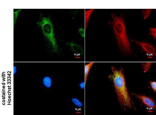
- IDH2 antibody detects IDH2 protein at Mitochondria by immunofluorescent analysis. Sample: HeLa cells were fixed in 4% paraformaldehyde/PBS for 15 min. Green: IDH2 protein stained by IDH2 antibody (Product # MA5-17271) diluted at 1:500. Red: alpha Tubulin protein stained by alpha Tubulin antibody (Product # PA5-85157) diluted at 1:500. Blue: Hoechst 33342 staining.

- Submitted by
- Invitrogen Antibodies (provider)
- Main image
- Experimental details
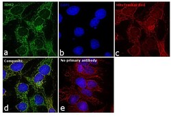
- Immunofluorescence analysis of IDH2 was performed using 70% confluent log phase Hep G2 cells. The cells were fixed with 4% paraformaldehyde for 10 minutes, permeabilized with 0.1% Triton™ X-100 for 15 minutes, and blocked with 1% BSA for 1 hour at room temperature. The cells were labeled with IDH2 Monoclonal Antibody (Product # MA5-17271) at 1:100 dilution in 0.1% BSA, incubated at 4 degree Celsius overnight and then labeled with Goat anti-Mouse IgG (H+L) Superclonal™ Secondary Antibody, Alexa Fluor® 488 conjugate (Product # A28175) at a dilution of 1:2000 for 45 minutes at room temperature (Panel a: green). Nuclei (Panel b: blue) were stained with SlowFade® Gold Antifade Mountant with DAPI (Product # S36938). Mitochondria (Panel c: red) was stained with Mitotracker Red CMXRos (Product # M7512). Panel d represents the merged image showing mitochondrial localization. Panel e represents control cells with no primary antibody to assess background. The images were captured at 60X magnification.

Supportive validation
- Submitted by
- Invitrogen Antibodies (provider)
- Main image

- Experimental details
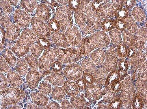
- Immunohistochemistry (Paraffin) analysis of IDH2 was performed in paraffin-embedded mouse kidney tissue using IDH2 Monoclonal Antibody (GT673) (Product # MA5-17271) at a dilution of 1:500.
- Submitted by
- Invitrogen Antibodies (provider)
- Main image
- Experimental details
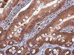
- Immunohistochemistry (Paraffin) analysis of IDH2 was performed in paraffin-embedded rat duodenum tissue using IDH2 Monoclonal Antibody (GT673) (Product # MA5-17271) at a dilution of 1:500.
