Explore
 Validate
 Learn
Learn Immunohistochemistry
Antibody data
- Antibody Data
- Antigen structure
- References [2]
- Comments [0]
- Validations
- Immunohistochemistry [1]
- Other assay [1]
Submit
Validation data
Reference
Comment
Report error
- Product number
- PA5-36089 - Provider product page

- Provider
- Invitrogen Antibodies
- Product name
- SENP7 Polyclonal Antibody
- Antibody type
- Polyclonal
- Antigen
- Synthetic peptide
- Description
- This antibody detects endogenous protein at a molecular weight of 112 kDa. Purity is >95% by SDS-PAGE.
- Reactivity
- Human, Mouse, Rat
- Host
- Rabbit
- Isotype
- IgG
- Vial size
- 100 μL
- Concentration
- 1 mg/mL
- Storage
- Store at 4°C short term. For long term storage, store at -20°C, avoiding freeze/thaw cycles.
Submitted references SENP7 senses oxidative stress to sustain metabolic fitness and antitumor functions of CD8+ T cells.
Extensive SUMO Modification of Repressive Chromatin Factors Distinguishes Pluripotent from Somatic Cells.
Wu Z, Huang H, Han Q, Hu Z, Teng XL, Ding R, Ye Y, Yu X, Zhao R, Wang Z, Zou Q
The Journal of clinical investigation 2022 Apr 1;132(7)
The Journal of clinical investigation 2022 Apr 1;132(7)
Extensive SUMO Modification of Repressive Chromatin Factors Distinguishes Pluripotent from Somatic Cells.
Theurillat I, Hendriks IA, Cossec JC, Andrieux A, Nielsen ML, Dejean A
Cell reports 2020 Sep 15;32(11):108146
Cell reports 2020 Sep 15;32(11):108146
No comments: Submit comment
Supportive validation
- Submitted by
- Invitrogen Antibodies (provider)
- Main image
- Experimental details
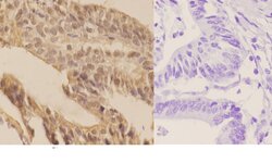
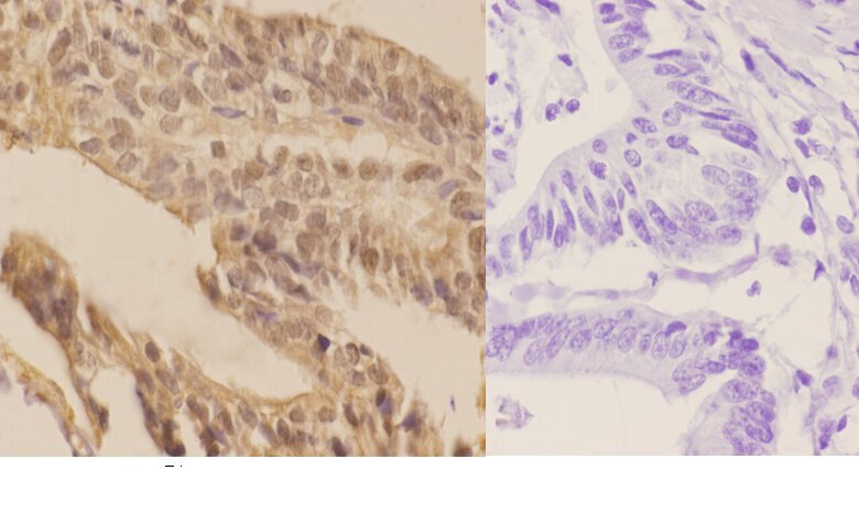
- Immunohistochemistry analysis of SENP7 in paraffin-embedded human colon carcinoma tissue (cytoplasmic and nucleus staining) and negative control (right, with PBS only). Samples were incubated with SENP7 polyclonal antibody (Product # PA5-36089) at a dilution of 1:50, followed by goat Anti-Rabbit IgG-biotin and avidin peroxidase.
Supportive validation
- Submitted by
- Invitrogen Antibodies (provider)
- Main image
- Experimental details
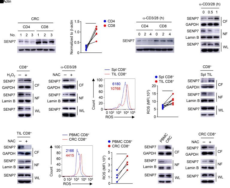
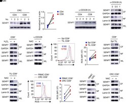
- Figure 1 ROS trigger cytosolic translocation of SENP7 in tumor-infiltrating CD8 + T cells. ( A and B ) Immunoblot analysis of the indicated proteins ( A ) and quantification of SENP7 expression ( B , n = 6) in CD4 + and CD8 + T cells from CRC tissues. ( C ) Immunoblot analysis of the indicated proteins in CD4 + and CD8 + T cells from WT mice stimulated with anti-CD3 and anti-CD28 antibodies (alpha-CD3/28). ( D - F ) Immunoblot analysis using whole-cell lysates (WL) and nuclear (NF) and cytoplasmic (CF) fractions of CD8 + T cells from WT mice stimulated with anti-CD3 and anti-CD28 antibodies ( D ), CD8 + T cells from WT mice treated with 0.2 mM H 2 O 2 for 1 hour ( E ), and CD8 + T cells from WT mice stimulated with anti-CD3 and anti-CD28 antibodies plus 10 mM NAC for 1 hour ( F ). ( G and H ) Histogram shows the MFI of ROS ( G ) and quantification of the MFI of ROS ( H , n = 7) in CD8 + T cells from the spleens (Spl) and tumors (TIL) of tumor-bearing mice (day 7 after injection of tumors with MC38 cells). ( I and J ) Immunoblot analysis using CD8 + T cells from the spleens and tumors of tumor-bearing mice ( I ) and tumor-infiltrating CD8 + T cells treated with 10 mM NAC for 1 hour ( J ). ( K and L ) Histogram shows the MFI of ROS ( K ) and quantification of the MFI of ROS ( L , n = 4) in CD8 + T cells from patient-derived PBMCs and CRC tissues. ( M and N ) Immunoblot analysis of the indicated proteins in CD8 + T cells from patient-derived PBMCs and CRC tissues ( M ) and CD8 +