 Explore
 Validate
 Learn
Learn Western blot
Antibody data
- Antibody Data
- Antigen structure
- References [6]
- Comments [0]
- Validations
- Western blot [8]
- Immunocytochemistry [1]
Submit
Validation data
Reference
Comment
Report error
- Product number
- PA1-31552 - Provider product page

- Provider
- Invitrogen Antibodies
- Product name
- TFEB Polyclonal Antibody
- Antibody type
- Polyclonal
- Antigen
- Synthetic peptide
- Description
- Expected to cross react with human due to sequence homology.
- Concentration
- 0.5 mg/mL
Submitted references Eicosapentaenoic acid attenuates renal lipotoxicity by restoring autophagic flux.
Sustained activation of autophagy suppresses adipocyte maturation via a lipolysis-dependent mechanism.
Mammalian Atg8 proteins and the autophagy factor IRGM control mTOR and TFEB at a regulatory node critical for responses to pathogens.
Transcription factor EB overexpression prevents neurodegeneration in experimental synucleinopathies.
AKT inhibition-mediated dephosphorylation of TFE3 promotes overactive autophagy independent of MTORC1 in cadmium-exposed bone mesenchymal stem cells.
Inhibiting ROS-TFEB-Dependent Autophagy Enhances Salidroside-Induced Apoptosis in Human Chondrosarcoma Cells.
Yamamoto T, Takabatake Y, Minami S, Sakai S, Fujimura R, Takahashi A, Namba-Hamano T, Matsuda J, Kimura T, Matsui I, Kaimori JY, Takeda H, Takahashi M, Izumi Y, Bamba T, Matsusaka T, Niimura F, Yanagita M, Isaka Y
Autophagy 2021 Jul;17(7):1700-1713
Autophagy 2021 Jul;17(7):1700-1713
Sustained activation of autophagy suppresses adipocyte maturation via a lipolysis-dependent mechanism.
Zhang X, Wu D, Wang C, Luo Y, Ding X, Yang X, Silva F, Arenas S, Weaver JM, Mandell M, Deretic V, Liu M
Autophagy 2020 Sep;16(9):1668-1682
Autophagy 2020 Sep;16(9):1668-1682
Mammalian Atg8 proteins and the autophagy factor IRGM control mTOR and TFEB at a regulatory node critical for responses to pathogens.
Kumar S, Jain A, Choi SW, da Silva GPD, Allers L, Mudd MH, Peters RS, Anonsen JH, Rusten TE, Lazarou M, Deretic V
Nature cell biology 2020 Aug;22(8):973-985
Nature cell biology 2020 Aug;22(8):973-985
Transcription factor EB overexpression prevents neurodegeneration in experimental synucleinopathies.
Arotcarena ML, Bourdenx M, Dutheil N, Thiolat ML, Doudnikoff E, Dovero S, Ballabio A, Fernagut PO, Meissner WG, Bezard E, Dehay B
JCI insight 2019 Aug 22;4(16)
JCI insight 2019 Aug 22;4(16)
AKT inhibition-mediated dephosphorylation of TFE3 promotes overactive autophagy independent of MTORC1 in cadmium-exposed bone mesenchymal stem cells.
Pi H, Li M, Zou L, Yang M, Deng P, Fan T, Liu M, Tian L, Tu M, Xie J, Chen M, Li H, Xi Y, Zhang L, He M, Lu Y, Chen C, Zhang T, Wang Z, Yu Z, Gao F, Zhou Z
Autophagy 2019 Apr;15(4):565-582
Autophagy 2019 Apr;15(4):565-582
Inhibiting ROS-TFEB-Dependent Autophagy Enhances Salidroside-Induced Apoptosis in Human Chondrosarcoma Cells.
Zeng W, Xiao T, Cai A, Cai W, Liu H, Liu J, Li J, Tan M, Xie L, Liu Y, Yang X, Long Y
Cellular physiology and biochemistry : international journal of experimental cellular physiology, biochemistry, and pharmacology 2017;43(4):1487-1502
Cellular physiology and biochemistry : international journal of experimental cellular physiology, biochemistry, and pharmacology 2017;43(4):1487-1502
No comments: Submit comment
Supportive validation
- Submitted by
- Invitrogen Antibodies (provider)
- Main image
- Experimental details
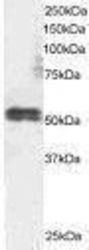
- Western blot analysis of TFEB using a TFEB polyclonal antibody (Product # PA1-31552).

- Submitted by
- Invitrogen Antibodies (provider)
- Main image
- Experimental details
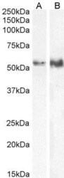
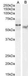
- Western Blot analysis of TFEB was performed by loading 35 µg (in RIPA buffer) of HeLa (A) and Jurkat (B) cell lysates. Proteins were transferred to a membrane and probed with a TFEB Polyclonal Antibody (Product # PA1-31552) at a dilution of 0.5 µg/mL.

- Submitted by
- Invitrogen Antibodies (provider)
- Main image
- Experimental details
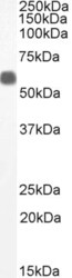
- Western Blot analysis of human thymus lysate (35 µg protein in RIPA buffer) using TFEB Polyclonal Antibody (Product # PA1-31552). Dilution: 1 µg/mL.

- Submitted by
- Invitrogen Antibodies (provider)
- Main image
- Experimental details
- Western Blot analysis of TFEB was performed by loading 35 µg (in RIPA buffer) of human thymus lysates. Proteins were transferred to a membrane and probed with a TFEB Polyclonal Antibody (Product # PA1-31552) at a dilution of 1 µg/mL.

- Submitted by
- Invitrogen Antibodies (provider)
- Main image
- Experimental details
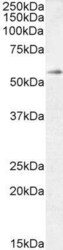
- Western Blot analysis of Daudi (Lane A) and HepG2 (Lane B) cell lysate (35 µg protein in RIPA buffer) using TFEB Polyclonal Antibody (Product # PA1-31552). Dilution: 0.3 µg/mL (Lane A) and 0.5 µg/mL (Lane B).

- Submitted by
- Invitrogen Antibodies (provider)
- Main image

- Experimental details
- Knockdown of TFEB was achieved by transfecting Raji with TFEB specific siRNAs (Silencer® select Product # s15495, s15496). Western blot analysis (Fig. a) was performed using whole cell extracts from the TFEB knockdown cells (lane 3), non-targeting scrambled siRNA transfected cells (lane 2) and untransfected cells (lane 1). The blot was probed with TFEB Polyclonal Antibody (Product # PA1-31552, 1 µg/mL ) and Rabbit anti-Goat IgG (H+L), Superclonal™ Recombinant Secondary Antibody, HRP (Product # A27014, 1:4000 dilution). Densitometric analysis of this western blot is shown in histogram (Fig. b). Decrease in signal upon siRNA mediated knock down confirms that antibody is specific to TFEB.

- Submitted by
- Invitrogen Antibodies (provider)
- Main image
- Experimental details
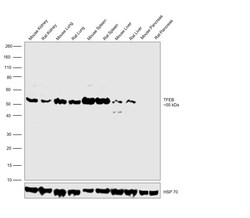
- Western blot was performed using Anti-TFEB Polyclonal Antibody (Product # PA1-31552) and a 55 kDa band corresponding to TFEB was observed across tissue tested except Mouse and Rat Pancreas. Tissue extracts of Mouse Kidney (Lane 1), Rat Kidney (Lane 2), Mouse Lung (Lane 3), Rat Lung (Lane 4), Mouse Spleen (Lane 5), Rat Spleen (Lane 6), Mouse Liver (Lane 7), Rat Liver (Lane 8), Mouse Pancreas (Lane 9) and Rat Pancreas (Lane 10) were electrophoresed using NuPAGE™ 4-12% Bis-Tris Protein Gel (Product # NP0322BOX). Resolved proteins were then transferred onto a nitrocellulose membrane (Product # IB23001) by iBlot® 2 Dry Blotting System (Product # IB21001). The blot was probed with the primary antibody (0.5 µg/mL) and detected by chemiluminescence with Rabbit anti-Goat IgG (H+L) Superclonal™ Recombinant Secondary Antibody, HRP (Product # A27014, 1:4000 dilution) using the iBright FL 1000 (Product # A32752). Chemiluminescent detection was performed using Novex® ECL Chemiluminescent Substrate Reagent Kit (Product # WP20005).

- Submitted by
- Invitrogen Antibodies (provider)
- Main image
- Experimental details
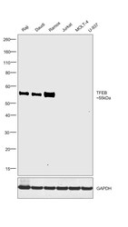
- Western blot was performed using Anti-TFEB Polyclonal Antibody (Product # PA1-31552) and a 55kDa band corresponding to TFEB was observed across all cell lines tested except Jurkat, MOLT-4 and U-937 which are known to have low TFEB expression (DOI: 10.1128/mcb.10.8.4384). Whole cell extracts (30 µg lysate) of Raji (Lane 1), Daudi (Lane 2), Ramos (Lane 3), Jurkat (Lane 4), MOLT-4 (Lane 5) and U-937 (Lane 6) were electrophoresed using NuPAGE™ 4-12% Bis-Tris Protein Gel (Product # NP0321BOX). Resolved proteins were then transferred onto a Nitrocellulose membrane (Product # IB23001) by iBlot® 2 Dry Blotting System (Product # IB21001). The blot was probed with the primary antibody (1 µg/mL dilution) and detected by chemiluminescence with Rabbit anti-Goat IgG (H+L), Superclonal™ Recombinant Secondary Antibody, HRP (Product # A27014, 1:4000 dilution) using the iBright FL 1000 (Product # A32752). Chemiluminescent detection was performed using Novex® ECL Reagent Kit (Product # WP20005).

Supportive validation
- Submitted by
- Invitrogen Antibodies (provider)
- Main image

- Experimental details
- Immunofluorescence analysis of TFEB was performed using 70% confluent log phase HEK-293 cells. The cells were fixed with 4% paraformaldehyde for 10 minutes, permeabilized with 0.1% Triton™ X-100 for 15 minutes, and blocked with 2% BSA for 1 hour at room temperature. The cells were labeled with TFEB Polyclonal Antibody (Product # PA1-31552) at 5 µg/mL in 0.1% BSA and incubated overnight at 4 degree and then labeled with Donkey anti-Goat IgG (H+L) Highly Cross-Adsorbed Secondary Antibody, Alexa Fluor Plus 488 conjugate (Product # A32814) at a dilution of 1:2000 for 45 minutes at room temperature (Panel a: green). Nuclei (Panel b: blue) were stained with ProLong™ Diamond Antifade Mountant with DAPI (Product # P36962). F-actin (Panel c: red) was stained with Rhodamine Phalloidin (Product # R415, 1:300). Panel d represents the composite image showing nuclear and cytoplasmic localization of TFEB in HEK-293 cells. Panel e represents control cells with no primary antibody to assess background. The images were captured at 60X magnification.
