Explore
 Validate
 Learn
Learn Western blot
Antibody data
- Antibody Data
- Antigen structure
- References [1]
- Comments [0]
- Validations
- Western blot [1]
- Immunocytochemistry [1]
Submit
Validation data
Reference
Comment
Report error
- Product number
- MAB9170-100 - Provider product page

- Provider
- R&D Systems
- Product name
- Human TFEB Antibody
- Antibody type
- Monoclonal
- Description
- Protein A or G purified from hybridoma culture supernatant. Detects human TFEB in direct ELISAs and Western blots.
- Reactivity
- Human
- Host
- Mouse
- Conjugate
- Unconjugated
- Antigen sequence
P19484- Isotype
- IgG
- Antibody clone number
- 954604
- Vial size
- 100 ug
- Storage
- Use a manual defrost freezer and avoid repeated freeze-thaw cycles. 12 months from date of receipt, -20 to -70 °C as supplied. 1 month, 2 to 8 °C under sterile conditions after reconstitution. 6 months, -20 to -70 °C under sterile conditions after reconstitution.
Submitted references IFNα Impairs Autophagic Degradation of mtDNA Promoting Autoreactivity of SLE Monocytes in a STING-Dependent Fashion.
Gkirtzimanaki K, Kabrani E, Nikoleri D, Polyzos A, Blanas A, Sidiropoulos P, Makrigiannakis A, Bertsias G, Boumpas DT, Verginis P
Cell reports 2018 Oct 23;25(4):921-933.e5
Cell reports 2018 Oct 23;25(4):921-933.e5
No comments: Submit comment
Supportive validation
- Submitted by
- R&D Systems (provider)
- Main image
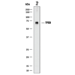
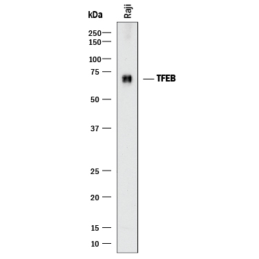
- Experimental details
- Detection of Human TFEB by Western Blot. Western blot shows lysates of Raji human Burkitt's lymphoma cell line. PVDF membrane was probed with 2 µg/mL of Mouse Anti-Human TFEB Monoclonal Antibody (Catalog # MAB9170) followed by HRP-conjugated Anti-Mouse IgG Secondary Antibody (Catalog # HAF018). A specific band was detected for TFEB at approximately 70 kDa (as indicated). This experiment was conducted under reducing conditions and using Immunoblot Buffer Group 1.
Supportive validation
- Submitted by
- R&D Systems (provider)
- Main image
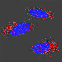
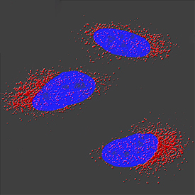
- Experimental details
- TFEB in A549 Human Cell Line. TFEB was detected in immersion fixed A549 human lung carcinoma cell line using Mouse Anti-Human TFEB Monoclonal Antibody (Catalog # MAB9170) at 3 µg/mL for 3 hours at room temperature. Cells were stained using the NorthernLights™ 557-conjugated Anti-Mouse IgG Secondary Antibody (red; Catalog # NL007) and counterstained with DAPI (blue). Specific staining was localized to cytoplasm. View our protocol for Fluorescent ICC Staining of Cells on Coverslips.