 Explore
 Validate
 Learn
Learn Western blot
Antibody data
- Antibody Data
- Antigen structure
- References [0]
- Comments [0]
- Validations
- Western blot [3]
- Immunocytochemistry [1]
- Immunohistochemistry [2]
Submit
Validation data
Reference
Comment
Report error
- Product number
- NBP2-30082 - Provider product page

- Provider
- Novus Biologicals
- Product name
- Rabbit Polyclonal PDK4 Antibody
- Antibody type
- Polyclonal
- Description
- Ammonium sulfate precipitation.
- Reactivity
- Human, Mouse
- Host
- Rabbit
- Isotype
- IgG
- Vial size
- 0.4 ml
- Concentration
- 0.75 mg/ml
- Storage
- Store at 4C short term. Aliquot and store at -20C long term. Avoid freeze-thaw cycles.
No comments: Submit comment
Supportive validation
- Submitted by
- Novus Biologicals (provider)
- Main image
- Experimental details
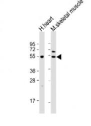
- Western Blot: PDK4 Antibody [NBP2-30082] - Anti-PDK4 Antibody at 1:2000 dilution Lane 1: human heart lysates Lane 2: mouse skeletal muscle lysates Lysates/proteins at 20 ug per lane. Secondary Goat Anti-Rabbit IgG, (H+L), Peroxidase conjugated at 1/10000 dilution Predicted band size : 46 kDa Blocking/Dilution buffer: 5% NFDM/TBST.

- Submitted by
- Novus Biologicals (provider)
- Main image

- Experimental details
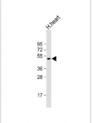
- Western Blot: PDK4 Antibody [NBP2-30082] - Anti-PDK4 Antibody (C-term) at 1:2000 dilution + human heart lysate Lysates/proteins at 20 ug per lane. Secondary Goat Anti-Rabbit IgG, (H+L), Peroxidase conjugated at 1/10000 dilution. Predicted band size : 46 kDa Blocking/Dilution buffer: 5% NFDM/TBST.
- Submitted by
- Novus Biologicals (provider)
- Main image
- Experimental details
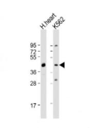
- Western Blot: PDK4 Antibody [NBP2-30082] - Anti-PDK4 Antibody (C-term) at 1:2000 dilution + human heart lysate Lysates/proteins at 20 ug per lane. Secondary Goat Anti-Rabbit IgG, (H+L), Peroxidase conjugated at 1/10000 dilution. Predicted band size : 46 kDa Blocking/Dilution buffer: 5% NFDM/TBST.

Supportive validation
- Submitted by
- Novus Biologicals (provider)
- Main image

- Experimental details
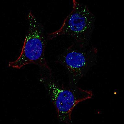
- Immunocytochemistry/Immunofluorescence: PDK4 Antibody [NBP2-30082] - Fluorescent confocal image of HeLa cells stained with PDK4 (C-term) antibody. HeLa cells were fixed with 4% PFA (20 min), permeabilized with Triton X-100 (0.2%, 30 min). Cells were then incubated with AP7041b PDK4 (C-term) primary antibody (1:100, 2 h at room temperature). For secondary antibody, Alexa Fluor 488 conjugated donkey anti-rabbit antibody (green) was used (1:1000, 1h). Nuclei were counterstained with Hoechst 33342 (blue) (10 ug/ml, 5 min). Note the highly specific localization of the PDK4 mainly to the cytoplasm.
Supportive validation
- Submitted by
- Novus Biologicals (provider)
- Main image
- Experimental details
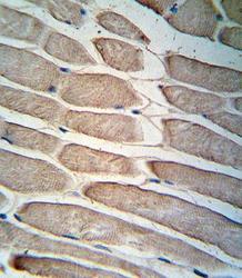
- Immunohistochemistry-Paraffin: PDK4 Antibody [NBP2-30082] - Formalin-fixed and paraffin-embedded human skeletal muscle reacted with (C-term) (NBP2-30082), which was peroxidase-conjugated to the secondary antibody, followed by DAB staining. This data demonstrates the use of this antibody for immunohistochemistry; clinical relevance has not been evaluated.

- Submitted by
- Novus Biologicals (provider)
- Main image
- Experimental details
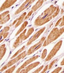
- Immunohistochemistry-Paraffin: PDK4 Antibody [NBP2-30082] - Human heart tissue sections by Immunohistochemistry (IHC-P - paraformaldehyde-fixed, paraffin-embedded sections). Tissue was fixed with formaldehyde and blocked with 3% BSA for 0. 5 hour at room temperature; antigen retrieval was by heat mediation with a citrate buffer (pH6). Samples were incubated with primary antibody (1/25) for 1 hours at 37 degrees C. A undiluted biotinylated goat polyvalent antibody was used as the secondary antibody.
