 Explore
 Validate
 Learn
Learn Western blot
Antibody data
- Antibody Data
- Antigen structure
- References [0]
- Comments [0]
- Validations
- Western blot [1]
- Immunocytochemistry [2]
- Immunohistochemistry [5]
Submit
Validation data
Reference
Comment
Report error
- Product number
- orb389347 - Provider product page

- Provider
- Biorbyt
- Product name
- FFAR1 antibody
- Antibody type
- Polyclonal
- Description
- Rabbit polyclonal antibody to FFAR1.
- Reactivity
- Mouse, Rat, Guinea Pig
- Host
- Rabbit
- Conjugate
- Unconjugated
- Isotype
- IgG
- Vial size
- 100 μg, 200 μg
- Concentration
- - 100 μg (in 200 μl): 0.5 mg/ml- 200 μg (in 400 μl): 0.5 mg/ml
- Storage
- Store at 4°C for up to two weeks. For long term storage, aliquot and store at -20°C, avoid freeze/thaw cycles.
No comments: Submit comment
Supportive validation
- Submitted by
- Biorbyt (provider)
- Main image

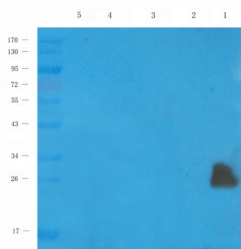
- Experimental details
- Western blot analysis of mouse pancreas (lane 1), rat ovary (lane 2), mouse spinal cord (lane 3), mouse brain (lane 4), mouse kidney (lane 5) using anti-FFAR1 (1 ug/ml)
Supportive validation
- Submitted by
- Biorbyt (provider)
- Main image
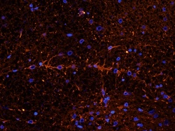
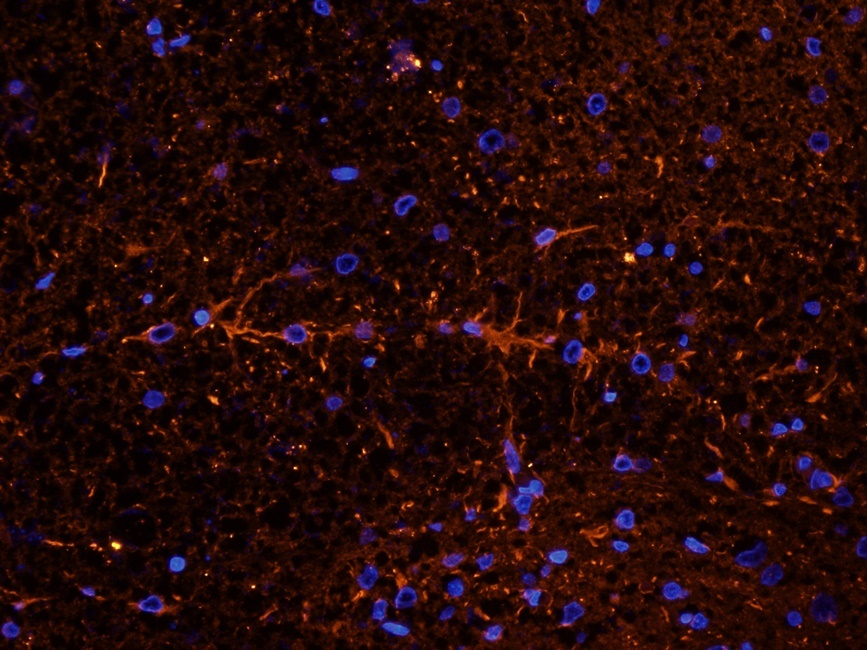
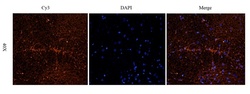
- Experimental details
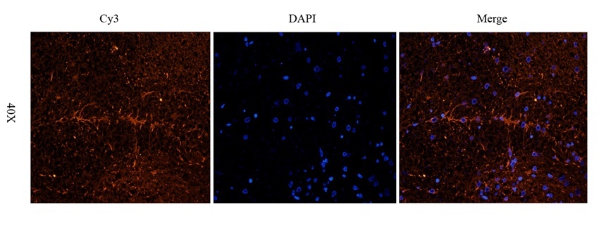
- Immunofluorescence analysis of rat spinal cord tissue using FFAR1 antibody (2.5 ug/ml)
- Submitted by
- Biorbyt (provider)
- Main image
- Experimental details
- IF analysis of rat spinal cord tissue using anti-FFAR1 (2.5 ug/ml)
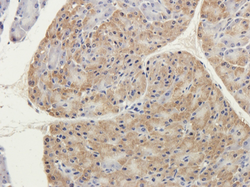
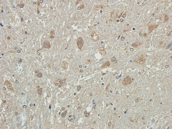
Supportive validation
- Submitted by
- Biorbyt (provider)
- Main image
- Experimental details
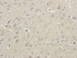
- IHC-P staining of guinea pig brain tissue using anti-FFAR1 (2.5 ug/ml)
- Submitted by
- Biorbyt (provider)
- Main image
- Experimental details
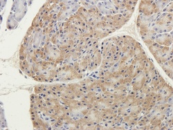
- IHC-P staining of guinea pig pancreas tissue using anti-FFAR1 (2.5 ug/ml)
- Submitted by
- Biorbyt (provider)
- Main image
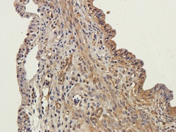
- Experimental details
- Immunohistochemical staining of paraffin embedded mouse ovary tissue using FFAR1 antibody (2.5 ug/ml)

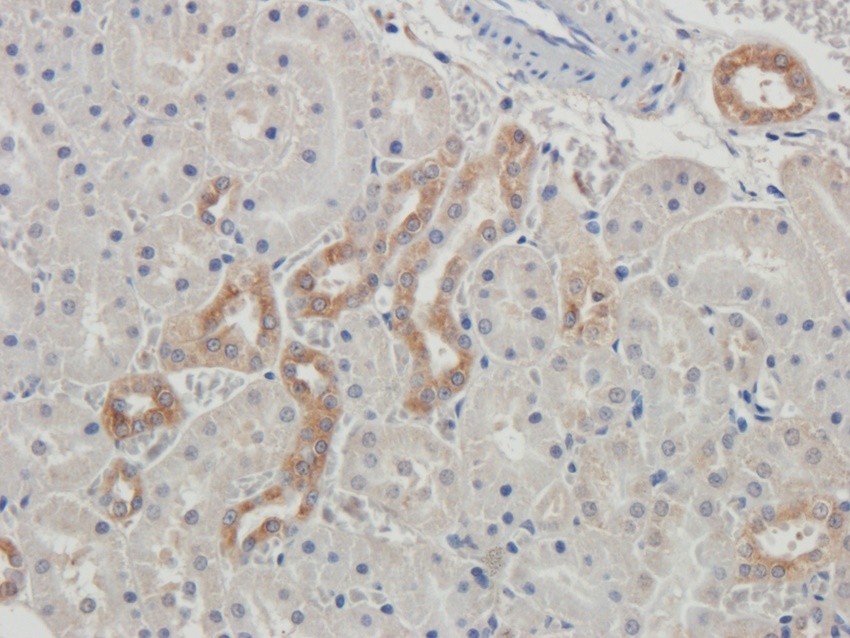
- Submitted by
- Biorbyt (provider)
- Main image
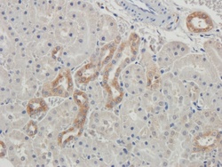
- Experimental details
- IHC-P staining of rat kidney tissue using anti-FFAR1 (2.5 ug/ml)
- Submitted by
- Biorbyt (provider)
- Main image
- Experimental details
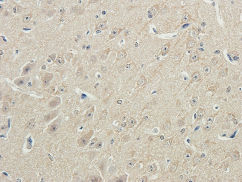
- IHC-P image of rat spinal cord tissue using anti-FFAR1 (2.5 ug/ml)
