Explore
 Validate
 Learn
Learn Western blot
Antibody data
- Antibody Data
- Antigen structure
- References [1]
- Comments [0]
- Validations
- Western blot [2]
- Immunohistochemistry [6]
Submit
Validation data
Reference
Comment
Report error
- Product number
- NBP1-91011 - Provider product page

- Provider
- Novus Biologicals
- Proper citation
- Novus Cat#NBP1-91011, RRID:AB_11023080
- Product name
- Rabbit Polyclonal Pepsinogen C/PGC/Progastricsin Antibody
- Antibody type
- Polyclonal
- Description
- Immunogen affinity purified. Specificity of human Pepsinogen C/PGC/Progastricsin antibody verified on a Protein Array containing target protein plus 383 other non-specific proteins.
- Reactivity
- Human
- Host
- Rabbit
- Isotype
- IgG
- Vial size
- 0.1 ml
- Storage
- Store at 4C short term. Aliquot and store at -20C long term. Avoid freeze-thaw cycles.
Submitted references Spatial maps of prostate cancer transcriptomes reveal an unexplored landscape of heterogeneity.
Berglund E, Maaskola J, Schultz N, Friedrich S, Marklund M, Bergenstråhle J, Tarish F, Tanoglidi A, Vickovic S, Larsson L, Salmén F, Ogris C, Wallenborg K, Lagergren J, Ståhl P, Sonnhammer E, Helleday T, Lundeberg J
Nature communications 2018 Jun 20;9(1):2419
Nature communications 2018 Jun 20;9(1):2419
No comments: Submit comment
Supportive validation
- Submitted by
- Novus Biologicals (provider)
- Main image
- Experimental details
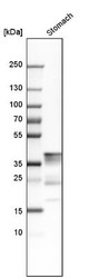
- Western Blot: Pepsinogen C/PGC/Progastricsin Antibody [NBP1-91011] - Analysis in human stomach tissue.

- Submitted by
- Novus Biologicals (provider)
- Main image
- Experimental details
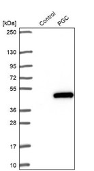
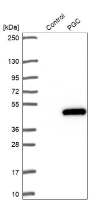
- Western Blot: Pepsinogen C/PGC/Progastricsin Antibody [NBP1-91011] - Analysis in control (vector only transfected HEK293T lysate) and PGC over-expression lysate (Co-expressed with a C-terminal myc-DDK tag (3.1 kDa) in mammalian HEK293T cells).
Supportive validation
- Submitted by
- Novus Biologicals (provider)
- Main image

- Experimental details
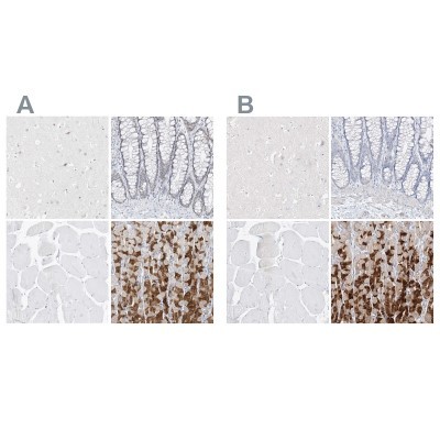
- Immunohistochemistry-Paraffin: Pepsinogen C/PGC/Progastricsin Antibody [NBP1-91011] - Staining of human cerebral cortex, colon, skeletal muscle and stomach using Anti-PGC antibody NBP1-91011 (A) shows similar protein distribution across tissues to independent antibody NBP1-91012 (B).
- Submitted by
- Novus Biologicals (provider)
- Main image
- Experimental details
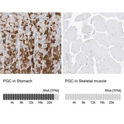
- Immunohistochemistry-Paraffin: Pepsinogen C/PGC/Progastricsin Antibody [NBP1-91011] - Analysis in human stomach and skeletal muscle tissues. Corresponding PGC RNA-seq data are presented for the same tissues.

- Submitted by
- Novus Biologicals (provider)
- Main image
- Experimental details
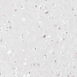
- Immunohistochemistry-Paraffin: Pepsinogen C/PGC/Progastricsin Antibody [NBP1-91011] - Staining of human cerebral cortex shows no positivity in neurons as expected.
- Submitted by
- Novus Biologicals (provider)
- Main image

- Experimental details
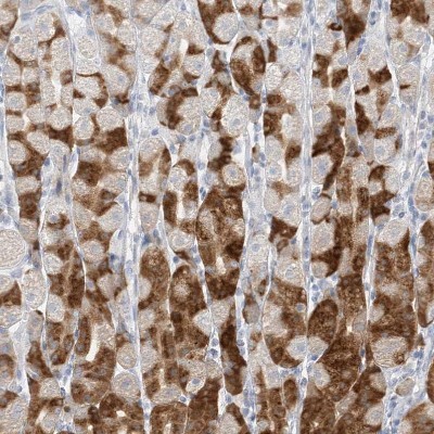
- Immunohistochemistry-Paraffin: Pepsinogen C/PGC/Progastricsin Antibody [NBP1-91011] - Staining of human stomach shows moderate cytoplasmic positivity in glandular cells.
- Submitted by
- Novus Biologicals (provider)
- Main image

- Experimental details
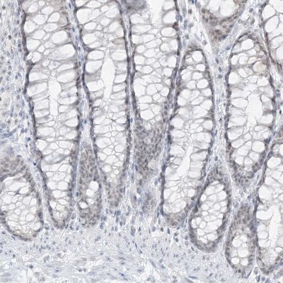
- Immunohistochemistry-Paraffin: Pepsinogen C/PGC/Progastricsin Antibody [NBP1-91011] - Staining of human colon shows low positivity in glandular cells as expected.
- Submitted by
- Novus Biologicals (provider)
- Main image
- Experimental details
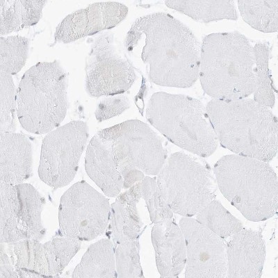
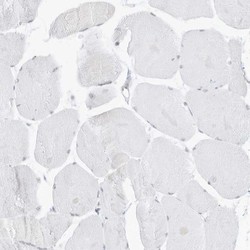
- Immunohistochemistry-Paraffin: Pepsinogen C/PGC/Progastricsin Antibody [NBP1-91011] - Staining of human skeletal muscle shows no positivity in myocytes as expected.