Explore
 Validate
 Learn
Learn Western blot
 Immunocytochemistry
Antibody data
- Antibody Data
- Antigen structure
- References [2]
- Comments [0]
- Validations
- Immunocytochemistry [2]
- Immunohistochemistry [2]
- Flow cytometry [2]
- Other assay [1]
Submit
Validation data
Reference
Comment
Report error
- Product number
- PA5-35340 - Provider product page

- Provider
- Invitrogen Antibodies
- Product name
- PIGR Polyclonal Antibody
- Antibody type
- Polyclonal
- Antigen
- Synthetic peptide
- Reactivity
- Human
- Host
- Rabbit
- Isotype
- IgG
- Vial size
- 400 μL
- Concentration
- 0.48 mg/mL
- Storage
- Store at 4°C short term. For long term storage, store at -20°C, avoiding freeze/thaw cycles.
Submitted references IgA transcytosis and antigen recognition govern ovarian cancer immunity.
A1 adenosine receptor signaling reduces Streptococcus pneumoniae adherence to pulmonary epithelial cells by targeting expression of platelet-activating factor receptor.
Biswas S, Mandal G, Payne KK, Anadon CM, Gatenbee CD, Chaurio RA, Costich TL, Moran C, Harro CM, Rigolizzo KE, Mine JA, Trillo-Tinoco J, Sasamoto N, Terry KL, Marchion D, Buras A, Wenham RM, Yu X, Townsend MK, Tworoger SS, Rodriguez PC, Anderson AR, Conejo-Garcia JR
Nature 2021 Mar;591(7850):464-470
Nature 2021 Mar;591(7850):464-470
A1 adenosine receptor signaling reduces Streptococcus pneumoniae adherence to pulmonary epithelial cells by targeting expression of platelet-activating factor receptor.
Bhalla M, Hui Yeoh J, Lamneck C, Herring SE, Tchalla EYI, Heinzinger LR, Leong JM, Bou Ghanem EN
Cellular microbiology 2020 Feb;22(2):e13141
Cellular microbiology 2020 Feb;22(2):e13141
No comments: Submit comment
Supportive validation
- Submitted by
- Invitrogen Antibodies (provider)
- Main image
- Experimental details
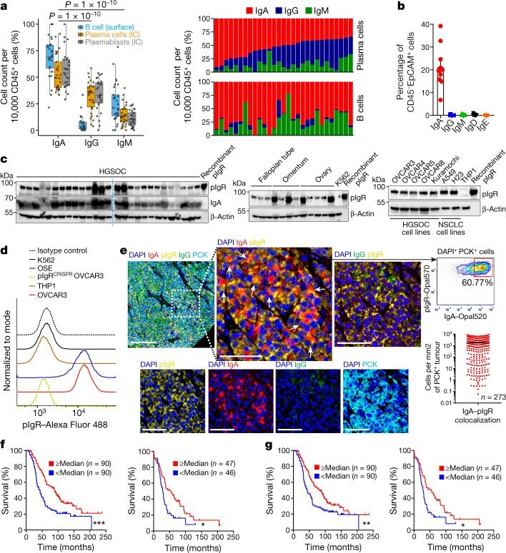
- Immunofluorescent analysis of PIGR in HepG2 cells using a PIGR polyclonal antibody (Product # PA5-35340) followed by detection using a fluorescent conjugated secondary antibody (green). Nuclei were stained with Dapi (blue).
- Submitted by
- Invitrogen Antibodies (provider)
- Main image

- Experimental details
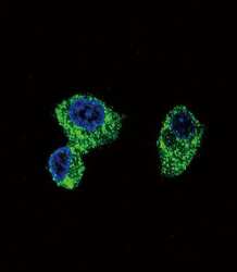
- Immunocytochemistry analysis of PIGR in HepG2 cells. Samples were incubated in PIGR polyclonal antibody (Product # PA5-35340) followed by Alexa Fluor 488-conjugated goat anti-rabbit lgG (green). DAPI was used to stain the cell nuclear (blue).

Supportive validation
- Submitted by
- Invitrogen Antibodies (provider)
- Main image

- Experimental details
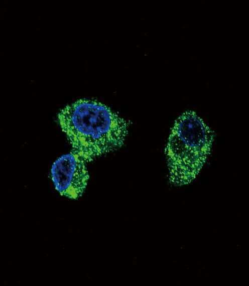
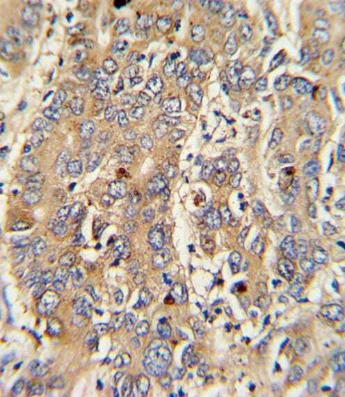
- Immunohistochemistry analysis of PIGR in formalin fixed and paraffin embedded human colon tissue. Samples were incubated with PIGR polyclonal antibody (Product # PA5-35340) followed by peroxidase conjugation of the secondary antibody and DAB staining. This data demonstrates the use of this antibody for immunohistochemistry. Clinical relevance has not been evaluated.
- Submitted by
- Invitrogen Antibodies (provider)
- Main image
- Experimental details
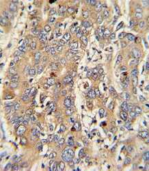
- Immunohistochemistry analysis of PIGR in formalin-fixed and paraffin-embedded human hepatocarcinoma. Samples were incubated with PIGR polyclonal antibody (Product # PA5-35340) which was peroxidase-conjugated to the secondary antibody, followed by DAB staining. This data demonstrates the use of this antibody for immunohistochemistry; clinical relevance has not been evaluated.
Supportive validation
- Submitted by
- Invitrogen Antibodies (provider)
- Main image
- Experimental details
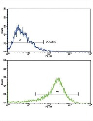
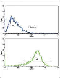
- Flow cytometry analysis of PIGR in HepG2 cells (bottom) compared to a negative control (top) using a PIGR polyclonal antibody (Product # PA5-35340) followed by detection using a FITC-conjugated goat-anti-rabbit secondary antibody.
- Submitted by
- Invitrogen Antibodies (provider)
- Main image
- Experimental details
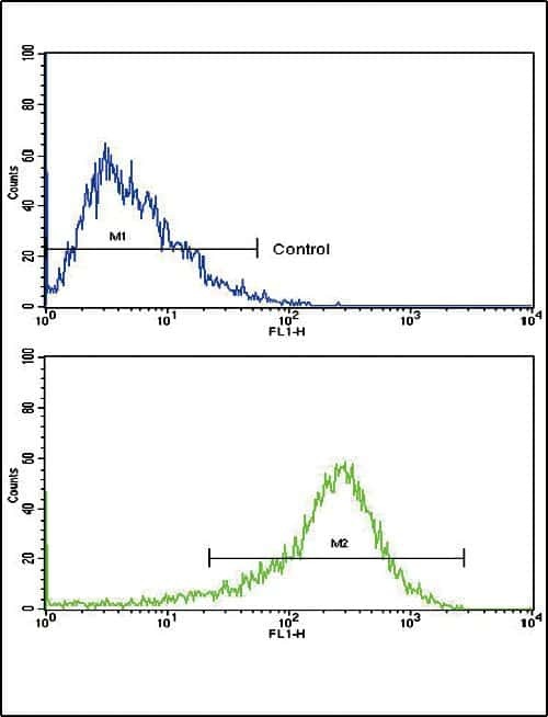
- Flow cytometry of PIGR in HepG2 cells (bottom histogram). Samples were incubated with PIGR polyclonal antibody (Product # PA5-35340) followed by FITC-conjugated goat-anti-rabbit secondary antibody. Negative control (top histogram).

Supportive validation
- Submitted by
- Invitrogen Antibodies (provider)
- Main image

- Experimental details
- Fig. 1 IgA-pIgR colocalization is associated with protective immunity in human ovarian cancer. a , Left, percentage of FACS cell counts of IgA + , IgG + or IgM + cells among Ig + B cells or plasmablasts or plasma cells, normalized to 10,000 viable CD45 + cells. B cells, CD45 + CD3 - CD19 + CD20 + cells; plasmablasts, CD45 + CD3 - CD19 + CD20 - CD38 high cells; plasma cells, CD45 + CD3 - CD19 + CD20 - CD138 + and CD45 + CD3 - CD19 - CD20 - CD138 + cells. Each dot represents one tumour ( n = 29). Details of box plots can be found in Methods. P values were obtained by a two-way analysis of variance (ANOVA) followed by Dunnett's test for multiple comparisons. Supplementary Table 1 provides further details on statistics. Right, bar graphs representing the percentage of each isotype produced by plasma cells (top) or B cells (bottom) in the same tumours, normalized to 10,000 viable CD45 + cells. IC, intracellular. b , IgA-coated CD45 - EpCAM + tumour epithelial cells (mean +- s.e.m., n = 10) in dissociated HGSOC. c , Expression of pIgR protein in independent HGSOC ( n = 27); tumour-free Fallopian tube ( n = 3), ovary ( n = 5) and omental ( n = 4) samples; ovarian tumour cell lines; and K562 leukaemia cells and THP1 monocyte cells (negative controls). Positive control, recombinant human pIgR. Western blots were repeated twice. NSCLC, non-small-cell lung cancer. d , Histograms showing FACS analysis of pIgR, in ovarian surface epithelial (OSE), K562, THP1, wild-type or PIGR -ablated (p