Explore
 Validate
 Learn
Learn Immunocytochemistry
Antibody data
- Antibody Data
- Antigen structure
- References [1]
- Comments [0]
- Validations
- Immunocytochemistry [2]
- Immunohistochemistry [1]
- Other assay [1]
Submit
Validation data
Reference
Comment
Report error
- Product number
- PA5-80323 - Provider product page

- Provider
- Invitrogen Antibodies
- Product name
- beta-2 Adrenergic Receptor Polyclonal Antibody
- Antibody type
- Polyclonal
- Antigen
- Synthetic peptide
- Description
- This product is preservative free. It is recommended to add sodium azide to avoid contamination (final concentration 0.05%-0.1%). This antibody has specificity for Human ADRB2.
- Reactivity
- Human
- Host
- Rabbit
- Isotype
- IgG
- Vial size
- 100 μL
- Concentration
- 1 mg/mL
- Storage
- Store at 4°C short term. For long term storage, store at -20°C, avoiding freeze/thaw cycles.
Submitted references Adrenergic activation modulates the signal from the Reissner fiber to cerebrospinal fluid-contacting neurons during development.
Cantaut-Belarif Y, Orts Del'Immagine A, Penru M, Pézeron G, Wyart C, Bardet PL
eLife 2020 Oct 13;9
eLife 2020 Oct 13;9
No comments: Submit comment
Supportive validation
- Submitted by
- Invitrogen Antibodies (provider)
- Main image
- Experimental details
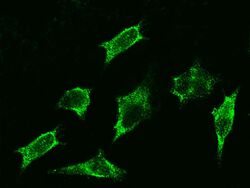
- Immunofluorescence staining of beta-2 Adrenergic Receptor in HeLa cells. Cells were fixed with 4% PFA, blocked with 10% serum, and incubated with beta-2 Adrenergic Receptor Polyclonal Antibody (Product # PA5-80323, 1:1,000) at 4°C overnight. Then cells were stained with the Alexa Fluor®488-conjugated Goat Anti-rabbit IgG secondary antibody (green). Positive staining was localized to cytoplasm.

- Submitted by
- Invitrogen Antibodies (provider)
- Main image

- Experimental details
- Immunofluorescence staining of beta-2 Adrenergic Receptor in HeLa cells. Cells were fixed with 4% PFA, blocked with 10% serum, and incubated with beta-2 Adrenergic Receptor Polyclonal Antibody (Product # PA5-80323, 1:1,000) at 4°C overnight. Then cells were stained with the Alexa Fluor®488-conjugated Goat Anti-rabbit IgG secondary antibody (green). Positive staining was localized to cytoplasm.

Supportive validation
- Submitted by
- Invitrogen Antibodies (provider)
- Main image
- Experimental details
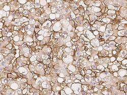
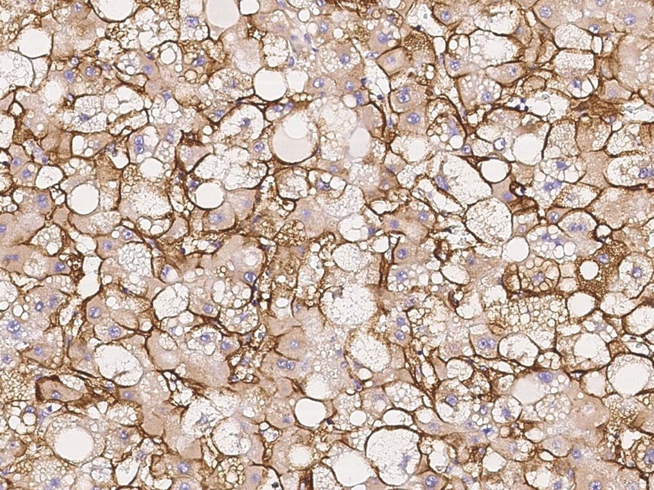
- Immunohistochemical staining of human beta-2 Adrenergic Receptor in human hepatoma with beta-2 Adrenergic Receptor Polyclonal Antibody (Product # PA5-80323, 1:1,000, formalin-fixed paraffin embedded sections).
Supportive validation
- Submitted by
- Invitrogen Antibodies (provider)
- Main image
- Experimental details
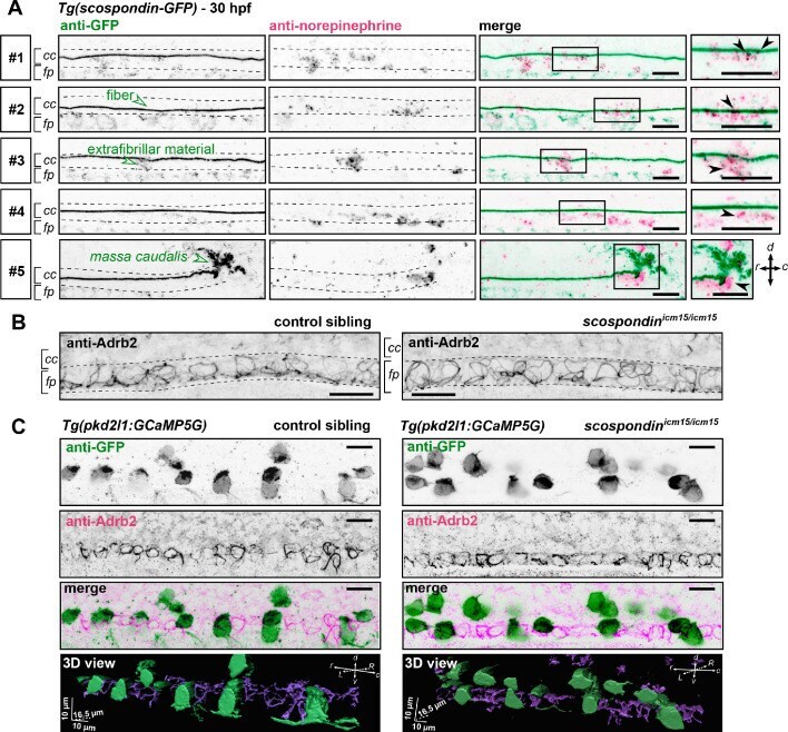
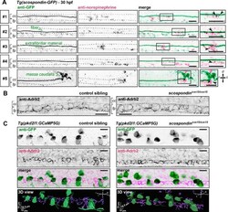
- Figure 6. The adrenergic receptor Adrb2 is expressed in cells ventral to the central canal in which norepinephrine can be detected. ( A ) Double immunodetection of GFP (left) and endogenous norepinephrine (middle) in the spinal cord of scospondin ut24Tg embryos at 30 hpf imaged laterally. Merged signals and close up of boxed regions are represented on the right. 5 representative examples (#1 to #5) out of 26 embryos are shown (no signal was detected in 6 embryos out of 26 in total, n = 2 independent experiments). In the central canal, norepinephrine positive signals can be detected as colocalized with the Reissner fiber itself (black arrowheads; embryos #1, 2, 3), with extrafibrillar material in the central canal (black arrowheads; embryos #4 and #5), and closely apposed to the massa caudalis located after the caudal limit of the central canal (black arrowhead; embryo #6). Scale bars: 10 um. r: rostral; c: caudal; d: dorsal; v: ventral; cc: central canal; fp: floor plate. ( B ) Immunohistochemistry for the adrenergic receptor Adrb2 in a 30 hpf control sibling (left, representative example out of 8 embryos) and a scospondin icm15/icm15 embryo (right, representative example out of 9 embryos). Adrb2 is distributed along the midline of the ventral most region of the neural tube (corresponding to the floor plate, fp) at the interface with the central canal (cc). Embryos are oriented rostral to the left and dorsal to the top. Scale bars: 10 um. ( C ) Sagittal views of double immuno