Explore
 Validate
 Learn
Learn Western blot
Antibody data
- Antibody Data
- Antigen structure
- References [0]
- Comments [0]
- Validations
- Western blot [1]
- Immunocytochemistry [1]
- Immunohistochemistry [1]
- Flow cytometry [1]
Submit
Validation data
Reference
Comment
Report error
- Product number
- AP52450PU-N - Provider product page

- Provider
- Acris Antibodies GmbH
- Proper citation
- Acris Antibodies GmbH Cat#AP52450PU-N, RRID:AB_11144075
- Product name
- anti LCAT (Center)
- Antibody type
- Polyclonal
- Antigen
- Synthetic peptide - KLH conjugated - corresponding to the central region (between 292-321aa) of human LCAT.
- Reactivity
- Human
- Host
- Rabbit
- Vial size
- 0.4 ml
- Concentration
- lot specific
No comments: Submit comment
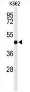
Supportive validation
- Submitted by
- Acris Antibodies GmbH (provider)
- Main image

- Experimental details
- Western blot analysis of LCAT (arrow) in K562 cell line lysates (35ug/lane) using LCAT antibody Cat.-No. AP52450PU-N.
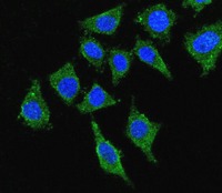
Supportive validation
- Submitted by
- Acris Antibodies GmbH (provider)
- Main image

- Experimental details
- Confocal immunofluorescent analysis with 293 cells using LCAT antibody Cat.-No. AP52450PU-N, followed by Alexa Fluor® 488-conjugated goat anti-rabbit lgG (green). DAPI was used to stain the cell nuclear (blue).
Supportive validation
- Submitted by
- Acris Antibodies GmbH (provider)
- Main image
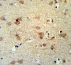
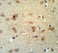
- Experimental details
- Immunohistochemistry analysis in brain tissue (Formalin-fixed, Paraffin-embedded) using LCAT antibody Cat.-No. AP52450PU-N, followed by peroxidase conjugation of the secondary antibody and DAB staining. This data demonstrates the use of the LCAT antibody for IHC; Clinical relevance has not been evaluated.
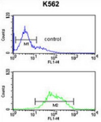
Supportive validation
- Submitted by
- Acris Antibodies GmbH (provider)
- Main image
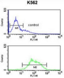
- Experimental details
- Flow cytometric analysis of K562 cells (bottom histogram) compared to a negative control cell (top histogram) using LCAT antibody Cat.-No. AP52450PU-N, followed by FITC-conjugated goat-anti-rabbit secondary antibodies.