Explore
 Validate
 Learn
Learn Western blot
Antibody data
- Antibody Data
- Antigen structure
- References [1]
- Comments [0]
- Validations
- Western blot [4]
- Immunocytochemistry [1]
- Immunoprecipitation [2]
- Immunohistochemistry [1]
Submit
Validation data
Reference
Comment
Report error
- Product number
- GTX111276 - Provider product page

- Provider
- GeneTex
- Proper citation
- GeneTex Cat#GTX111276, RRID:AB_1950216
- Product name
- Her4 / ErbB4 antibody [C1C3]
- Antibody type
- Polyclonal
- Reactivity
- Human, Mouse, Rat
- Host
- Rabbit
Submitted references The significance of Her2 on androgen receptor protein stability in the transition of androgen requirement in prostate cancer cells.
Hsu FN, Yang MS, Lin E, Tseng CF, Lin H
American journal of physiology. Endocrinology and metabolism 2011 May;300(5):E902-8
American journal of physiology. Endocrinology and metabolism 2011 May;300(5):E902-8
No comments: Submit comment
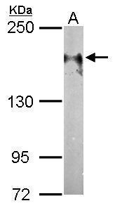
Supportive validation
- Submitted by
- GeneTex (provider)
- Main image

- Experimental details
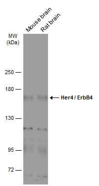
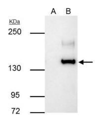
- Sample (50 ?g of whole cell lysate) A: Mouse brain 5% SDS PAGE GTX111276 diluted at 1:1000 The HRP-conjugated anti-rabbit IgG antibody (GTX213110-01) was used to detect the primary antibody.
- Submitted by
- GeneTex (provider)
- Main image
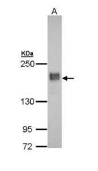
- Experimental details
- Sample (30 ?g of whole cell lysate) A: A431 (GTX27909) 5% SDS PAGE GTX111276 diluted at 1:5000 The HRP-conjugated anti-rabbit IgG antibody (GTX213110-01) was used to detect the primary antibody.

- Submitted by
- GeneTex (provider)
- Main image
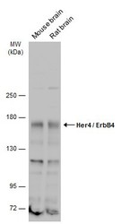
- Experimental details
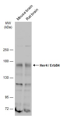
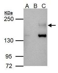
- Various tissue extracts (50 ?g) were separated by 5% SDS-PAGE, and the membrane was blotted with Her4 / ErbB4 antibody (GTX111276) diluted at 1:1000. The HRP-conjugated anti-rabbit IgG antibody (GTX213110-01) was used to detect the primary antibody.
- Submitted by
- GeneTex (provider)
- Main image
- Experimental details
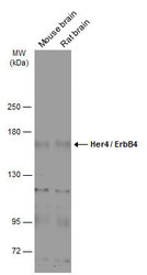
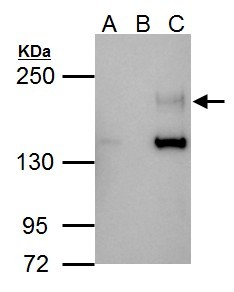
- Various tissue extracts (50 ?g) were separated by 5% SDS-PAGE, and the membrane was blotted with Her4 / ErbB4 antibody [C1C3] (GTX111276) diluted at 1:1000. The HRP-conjugated anti-rabbit IgG antibody (GTX213110-01) was used to detect the primary antibody.
Supportive validation
- Submitted by
- GeneTex (provider)
- Main image
- Experimental details
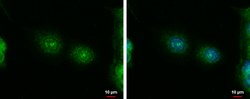
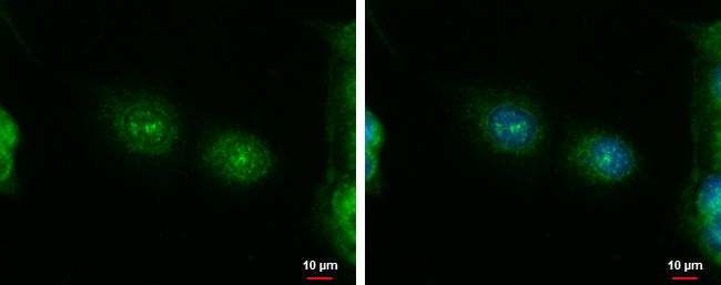
- ERBB4 antibody [C1C3] detects ERBB4 protein at cytoplasm and nucleus by immunofluorescent analysis.Sample: MCF-7 cells were fixed in ice-cold MeOH for 5 min.Green: ERBB4 protein stained by ERBB4 antibody [C1C3] (GTX111276) diluted at 1:500.Blue: Hoechst 33342 staining.
Supportive validation
- Submitted by
- GeneTex (provider)
- Main image
- Experimental details
- ERBB4 antibody [C1C3] immunoprecipitates ERBB4 protein in IP experiments.IP samples: MCF-7 whole cell extractA. Control with 4 £gg of preimmune Rabbit IgGB. Immunoprecipitation of ERBB4 protein by 4 £gg ERBB4 antibody [C1C3] (GTX111276)5 % SDS-PAGEThe immunoprecipitated ERBB4 protein was detected by ERBB4 antibody [C1C3] (GTX111276) diluted at 1:500.[EasyBlot anti-rabbit IgG (GTX221666-01) was used as a secondary reagent]

- Submitted by
- GeneTex (provider)
- Main image
- Experimental details
- Immunoprecipitation of Her4 / ErbB4 protein from MCF-7 whole cell extract using 4.5 ?g of Her4 / ErbB4 antibody [C1C3] (GTX111276).Western blot analysis was performed using Her4 / ErbB4 antibody [C1C3] (GTX111276) and diluted at 1:500.EasyBlot HRP-conjugated anti rabbit IgG antibody (GTX221666-01) was used to detect the primary antibody.
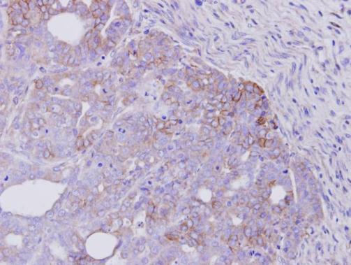
Supportive validation
- Submitted by
- GeneTex (provider)
- Main image
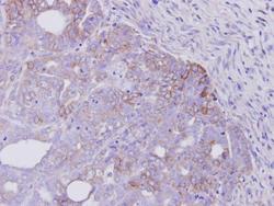
- Experimental details
- Immunohistochemical analysis of paraffin-embedded NCIN87 xenograft, using ERBB4(v-erb-A)(GTX111276) antibody at 1:500 dilution.