Explore
 Validate
 Learn
Learn Western blot
 Immunoprecipitation
Antibody data
- Antibody Data
- Antigen structure
- References [0]
- Comments [0]
- Validations
- Western blot [1]
- Immunocytochemistry [3]
- Immunohistochemistry [3]
- Flow cytometry [3]
Submit
Validation data
Reference
Comment
Report error
- Product number
- GTX80811 - Provider product page

- Provider
- GeneTex
- Proper citation
- GeneTex Cat#GTX80811, RRID:AB_625545
- Product name
- Her4 / ErbB4 antibody [HFR1]
- Antibody type
- Monoclonal
- Reactivity
- Human, Mouse
- Host
- Mouse
No comments: Submit comment
Supportive validation
- Submitted by
- GeneTex (provider)
- Main image

- Experimental details
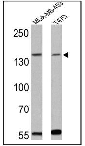
- WB analysis of MDA-MB-453 and T47D lysates (25 ug per lane) using ErbB4 antibody [HFR1] at a dilution of 1:300.
Supportive validation
- Submitted by
- GeneTex (provider)
- Main image
- Experimental details
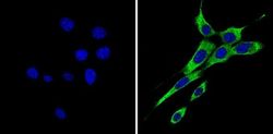
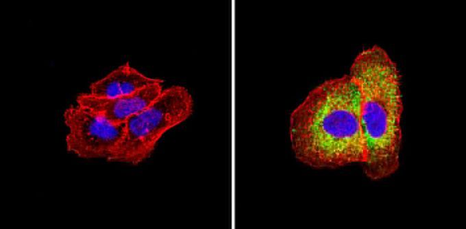
- ICC/IF analysis of NIH-3T3 cells with (right) or without (left) ErbB4 antibody [HFR1] at a dilution of 1:50 (green). Nuclei were stained with DAPI (blue). Images were taken at a magnification of 60x.

- Submitted by
- GeneTex (provider)
- Main image
- Experimental details
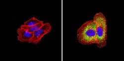
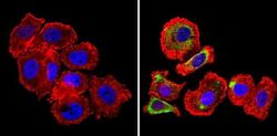
- ICC/IF analysis of A431 cells with (right) or without (left) ErbB4 antibody [HFR1] at a dilution of 1:50 (green). Actin was stained using Alexa Fluor 554 (red) and nuclei were stained with DAPI (blue). Images were taken at a magnification of 60x.
- Submitted by
- GeneTex (provider)
- Main image
- Experimental details
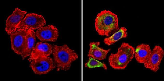
- ICC/IF analysis of SK-BR-3 cells with (right) or without (left) ErbB4 antibody [HFR1] at a dilution of 1:50 (green). Actin was stained using Alexa Fluor 554 (red) and nuclei were stained with DAPI (blue). Images were taken at a magnification of 60x.
Supportive validation
- Submitted by
- GeneTex (provider)
- Main image
- Experimental details
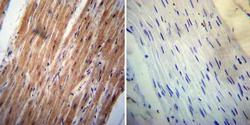
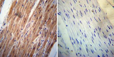
- IHC-P analysis of human heart tissue with (left) or without (right) ErbB4 antibody [HFR1] at a dilution of 1:200. To expose target proteins, heat induced antigen retrieval was performed using 10mM sodium citrate (pH6.0) buffer, microwaved for 8-15 minutes.
- Submitted by
- GeneTex (provider)
- Main image

- Experimental details
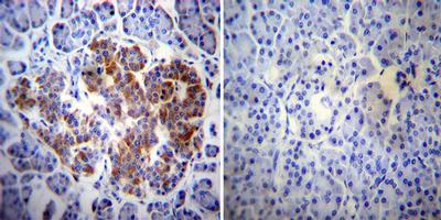
- IHC-P analysis of human pancreas tissue with (left) or without (right) ErbB4 antibody [HFR1] at a dilution of 1:200. To expose target proteins, heat induced antigen retrieval was performed using 10mM sodium citrate (pH6.0) buffer, microwaved for 8-15 minutes.
- Submitted by
- GeneTex (provider)
- Main image
- Experimental details
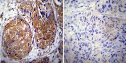
- IHC-P analysis of human breast carcinoma with (left) or without (right) ErbB4 antibody [HFR1] at a dilution of 1:20. To expose target proteins, heat induced antigen retrieval was performed using 10mM sodium citrate (pH6.0) buffer, microwaved for 8-15 minutes.

Supportive validation
- Submitted by
- GeneTex (provider)
- Main image
- Experimental details
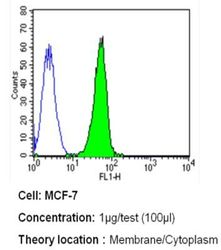
- FACS analysis of MCF-7 cells with ErbB4 antibody [HFR1] at a dilution of 1ug/test (green) or an isotype control (blue).

- Submitted by
- GeneTex (provider)
- Main image
- Experimental details
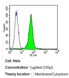
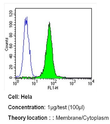
- FACS analysis of HeLa cells with ErbB4 antibody [HFR1] at a dilution of 1ug/test (green) or an isotype control (blue).
- Submitted by
- GeneTex (provider)
- Main image

- Experimental details
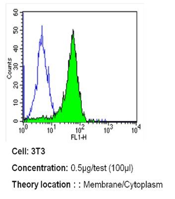
- FACS analysis of NIH-3T3 cells with ErbB4 antibody [HFR1] at a dilution of 0.5ug/test (green) or an isotype control (blue).