 Explore
 Validate
 Learn
Learn Western blot
 Immunohistochemistry
Antibody data
- Antibody Data
- Antigen structure
- References [4]
- Comments [0]
- Validations
- Immunohistochemistry [1]
Submit
Validation data
Reference
Comment
Report error
- Product number
- 14-9687-80 - Provider product page

- Provider
- Invitrogen Antibodies
- Product name
- ErbB4 (Her4) Monoclonal Antibody (HFR1), eBioscience™
- Antibody type
- Monoclonal
- Antigen
- Other
- Description
- Description: The monoclonal antibody HFR1 recognizes human and mouse epidermal growth factor receptor 4 (ErbB4 or HER4). ErbB4 belongs to the family of receptor tyrosine kinases (RTK) of which HER1 (EGRF), HER2 (ErbB2), and HER3 (ErbB3) are members. RTKs function to affect a variety of cell processes including proliferation, differentiation, migration, survival and apoptosis and are often disregulated in cancer. The ErbB4 protein contains extracellular ligand binding domains, a transmembrane region, and a cytoplasmic tail containing the tyrosine kinase domain and the binding site for the HFR1 antibody. Upon ligand binding, ErbB4 homo- or hetero-dimerizes with other family members, becomes phosphorylated, and can be cleaved allowing translocation to the nucleus. The HRF1 antibody recognizes membrane-bound, cytoplasmic and nuclear ErbB4. Cytosolic ErbB4 expression was found to correlate with a positive prognosis in a subset of breast cancer patients, while nuclear ErbB4 expression was inversely correlated with tumor grade and aggressiveness of breast tumors. Applications Reported: This HFR1 antibody has been reported for use in immunoprecipitation, western blotting, immunohistochemical staining of formalin-fixed paraffin embedded tissue sections, and immunocytochemistry. Applications Tested: This HFR1 antibody has been tested by immunocytochemistry on fixed and permeablilized A431 cells and can be used at less than or equal to 10 µg/mL. The HFR1 antibody has also been tested by immunohistochemistry on human FFPE tissue with low pH antigen retrieval and can be used at less than or equal to 10 µg/mL. It is recommended that the antibody be carefully titrated for optimal performance in the assay of interest. Purity: Greater than 90%, as determined by SDS-PAGE. Aggregation: Less than 10%, as determined by HPLC. Filtration: 0.2 µm post-manufacturing filtered.
- Reactivity
- Human, Mouse
- Host
- Mouse
- Isotype
- IgG
- Antibody clone number
- HFR1
- Vial size
- 25 μg
- Concentration
- 0.5 mg/mL
- Storage
- 4°C
Submitted references Subcellular localization of the HER4 intracellular domain, 4ICD, identifies distinct prognostic outcomes for breast cancer patients.
HER4 in breast cancer: comparison of antibodies against intra- and extra-cellular domains of HER4.
Nuclear expression of the c-erbB-4/HER-4 growth factor receptor in invasive breast cancers.
Expression of the c-erbB-3/HER-3 and c-erbB-4/HER-4 growth factor receptors and their ligands, neuregulin-1 alpha, neuregulin-1 beta, and betacellulin, in normal endometrium and endometrial cancer.
Thor AD, Edgerton SM, Jones FE
The American journal of pathology 2009 Nov;175(5):1802-9
The American journal of pathology 2009 Nov;175(5):1802-9
HER4 in breast cancer: comparison of antibodies against intra- and extra-cellular domains of HER4.
Tovey SM, Dunne B, Witton CJ, Cooke TG, Bartlett JM
Breast cancer research : BCR 2006;8(2):R19
Breast cancer research : BCR 2006;8(2):R19
Nuclear expression of the c-erbB-4/HER-4 growth factor receptor in invasive breast cancers.
Srinivasan R, Gillett CE, Barnes DM, Gullick WJ
Cancer research 2000 Mar 15;60(6):1483-7
Cancer research 2000 Mar 15;60(6):1483-7
Expression of the c-erbB-3/HER-3 and c-erbB-4/HER-4 growth factor receptors and their ligands, neuregulin-1 alpha, neuregulin-1 beta, and betacellulin, in normal endometrium and endometrial cancer.
Srinivasan R, Benton E, McCormick F, Thomas H, Gullick WJ
Clinical cancer research : an official journal of the American Association for Cancer Research 1999 Oct;5(10):2877-83
Clinical cancer research : an official journal of the American Association for Cancer Research 1999 Oct;5(10):2877-83
No comments: Submit comment
Supportive validation
- Submitted by
- Invitrogen Antibodies (provider)
- Main image
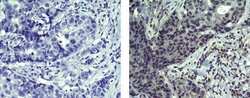
- Experimental details
- Immunohistochemistry of formalin-fixed paraffin embedded human infiltrating ductal carcinoma using 10 µg/mL Mouse IgG2b K Isotype Control Purified (left) or 10 µg/mL Anti-Human/Mouse ErbB4 (Her4) Purified (right) followed by Anti-Mouse IgG Biotin and DAB visualization.Nuclei are counterstained with hematoxylin.
