Explore
 Validate
 Learn
Learn Immunohistochemistry
Antibody data
- Antibody Data
- Antigen structure
- References [1]
- Comments [0]
- Validations
- Immunohistochemistry [3]
- Other assay [1]
Submit
Validation data
Reference
Comment
Report error
- Product number
- PA5-110928 - Provider product page

- Provider
- Invitrogen Antibodies
- Product name
- GPR182 Polyclonal Antibody
- Antibody type
- Polyclonal
- Antigen
- Recombinant protein fragment
- Description
- Immunogen sequence: LSPHFRGRLL NAVVHYLPKD QTKAGTCASS SSCSTQHSII ITKGDSQPAA AAPHPEPSLS FQAHHLLPNT SPISPTQPLT PS
- Reactivity
- Human
- Host
- Rabbit
- Isotype
- IgG
- Vial size
- 100 μL
- Storage
- Store at 4°C short term. For long term storage, store at -20°C, avoiding freeze/thaw cycles.
Submitted references GPR182 limits antitumor immunity via chemokine scavenging in mouse melanoma models.
Torphy RJ, Sun Y, Lin R, Caffrey-Carr A, Fujiwara Y, Ho F, Miller EN, McCarter MD, Lyons TR, Schulick RD, Kedl RM, Zhu Y
Nature communications 2022 Jan 10;13(1):97
Nature communications 2022 Jan 10;13(1):97
No comments: Submit comment
Supportive validation
- Submitted by
- Invitrogen Antibodies (provider)
- Main image
- Experimental details
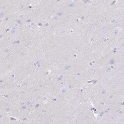
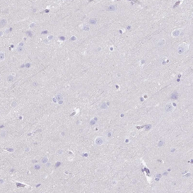
- Immunohistochemical analysis of GPR182 in human cerebral cortex using GPR182 Polyclonal Antibody (Product # PA5-110928) shows low expression as expected.
- Submitted by
- Invitrogen Antibodies (provider)
- Main image
- Experimental details
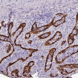
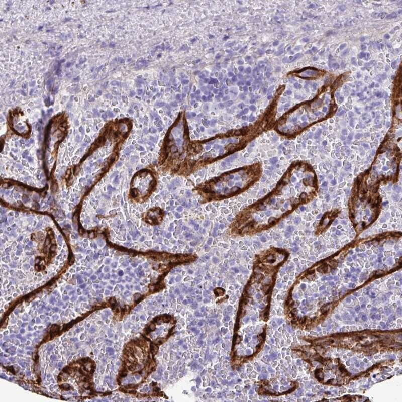
- Immunohistochemical analysis of GPR182 in human spleen using GPR182 Polyclonal Antibody (Product # PA5-110928) shows high expression.
- Submitted by
- Invitrogen Antibodies (provider)
- Main image
- Experimental details
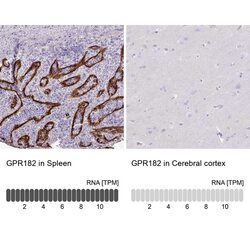
- Immunohistochemical analysis of GPR182 in human spleen and cerebral cortex tissues using GPR182 Polyclonal Antibody (Product # PA5-110928) using Anti-GPR182 antibody. Corresponding GPR182 RNA-seq data are presented for the same tissues.

Supportive validation
- Submitted by
- Invitrogen Antibodies (provider)
- Main image
- Experimental details
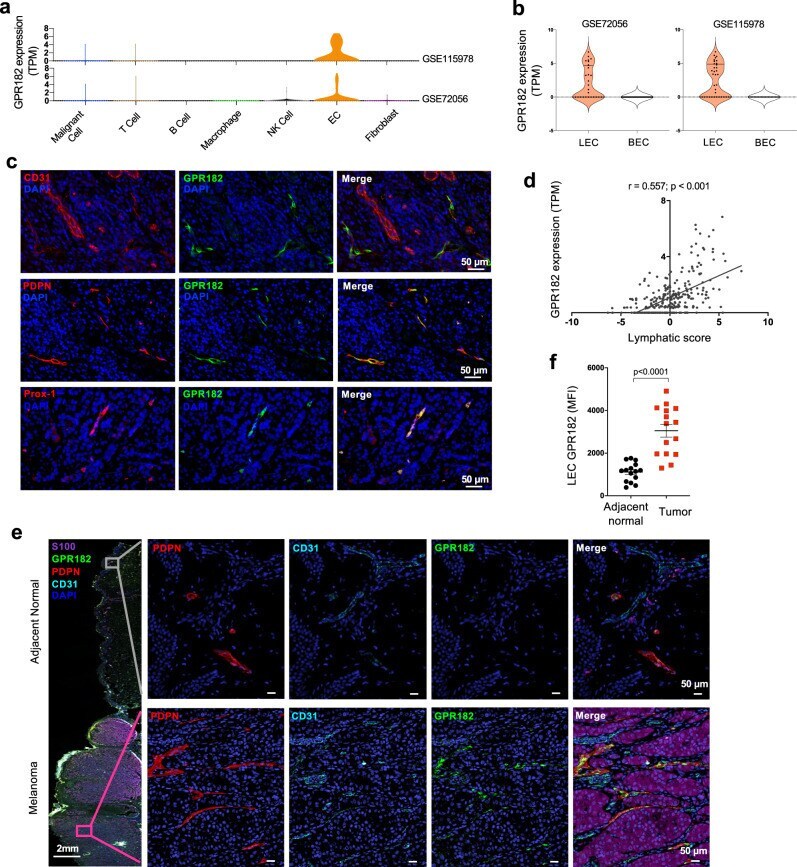
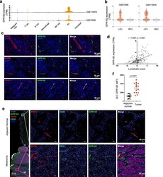
- Fig. 1 GPR182 is expressed by lymphatic endothelial cells in human melanoma. a , b Two published single-cell RNA sequencing datasets of human melanoma were queried for GPR182 expression based on cell type ( a ). b Analysis of ECs in human melanoma further revealed that GPR182 is primarily expressed in LECs. c Human melanoma tissues were stained for GPR182 (green) together with EC markers, including CD31, podoplanin (PDPN), or Prox-1. d A lymphatic score was generated using 288 metastatic melanoma samples from the TCGA database. The lymphatic score was calculated based on relative expression levels of PDPN , LYVE1 , and VEGFC in each sample. Lymphatic score was plotted against GPR182 mRNA expression level (RSEM, log2 normalized). Pearson's correlation coefficient, r , and p -values shown from two-sided test. e , f Human melanoma tissues with adjacent normal skin were stained for PDPN (red), CD31(blue), and GPR182 (green) (E); S-100 staining (brown) was used to identify melanocytes and tumor cells. f Quantification of GPR182 median fluorescent intensity (MFI) in PDPN+ lymphatic vessels from paired tumor and adjacent normal tissue. n = 15, P -value from two-sided paired t -test. Error bars represent SEM.