Explore
 Validate
 Learn
Learn Western blot
 Immunocytochemistry
Antibody data
- Antibody Data
- Antigen structure
- References [0]
- Comments [0]
- Validations
- Immunocytochemistry [4]
- Immunoprecipitation [1]
- Other assay [1]
Submit
Validation data
Reference
Comment
Report error
- Product number
- 703744 - Provider product page

- Provider
- Invitrogen Antibodies
- Product name
- PRMT3 Recombinant Rabbit Monoclonal Antibody (23H2L13)
- Antibody type
- Monoclonal
- Antigen
- Other
- Description
- This antibody is predicted to react with Mouse, Bovine.
- Reactivity
- Human, Rat
- Host
- Rabbit
- Isotype
- IgG
- Antibody clone number
- 23H2L13
- Vial size
- 100 μg
- Concentration
- 0.5 mg/mL
- Storage
- Store at 4°C short term. For long term storage, store at -20°C, avoiding freeze/thaw cycles.
No comments: Submit comment
Supportive validation
- Submitted by
- Invitrogen Antibodies (provider)
- Main image

- Experimental details
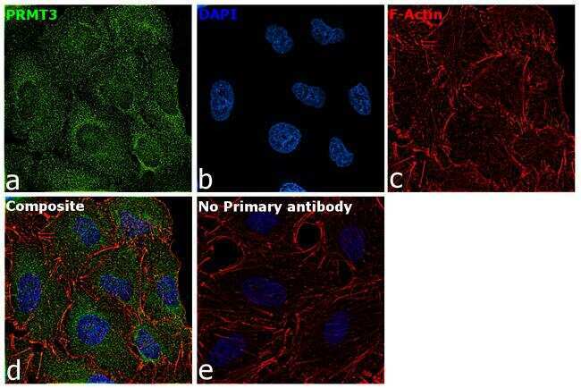
- For immunocytochemistry analysis, A549 cells were fixed and permeabilized for detection of endogenous PRMT3 using Anti-PRMT3 Recombinant Rabbit Monoclonal Antibody (Product # 703744) at a 1:100 dilution and labeled with Goat anti-Rabbit IgG (H+L) Superclonal™ Secondary Antibody, Alexa Fluor® 488 conjugate (Product # A27034) at a 1:2000 dilution. Panel a) shows representative cells that were stained for detection and localization of PRMT3 protein (green), Panel b) is stained for nuclei (blue) using ProLong™ Diamond Antifade Mountant with DAPI (Product # P36962). Panel c) represents cytoskeletal F-actin staining using Rhodamine Phalloidin (Product # R415) at a 1:300 dilution. Panel d) is a composite image of Panels a, b and c clearly demonstrating cytoplasmic localization of PRMT3. Panel e) represents control cells with no primary antibody to assess background. The images were captured at 60X magnification.
- Submitted by
- Invitrogen Antibodies (provider)
- Main image

- Experimental details
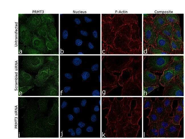
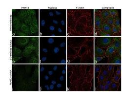
- Knockdown of PRMT3 was achieved by transfecting A549 cells with specific validated siRNA (Silencer® select Product # s19870). Immunofluorescence analysis was performed on A549 cells (untransfected, panel a-d), transfected with PRMT3 specific siRNA (panel i-l) or non-specific scrambled siRNA (panels e-h). Cells were fixed, permeabilized, and labeled with Anti-PRMT3 Recombinant Rabbit Monoclonal Antibody (Product # 703744) at a 1:100 dilution, followed by Goat anti-Rabbit IgG (H+L) Superclonal™ Secondary Antibody, Alexa Fluor® 488 conjugate (Product # A27034) at a 1:2000 dilution. Nuclei (blue) were stained using ProLong™ Diamond Antifade Mountant with DAPI (Product # P36962), and Rhodamine Phalloidin (Product # R415) at a 1:300 dilution was used for cytoskeletal F-actin (red) staining. Significant reduction of the signal was observed upon siRNA mediated knockdown (panel i-l) confirming the specificity of the antibody to PRMT3 (green). The images were captured at 60X magnification.
- Submitted by
- Invitrogen Antibodies (provider)
- Main image

- Experimental details
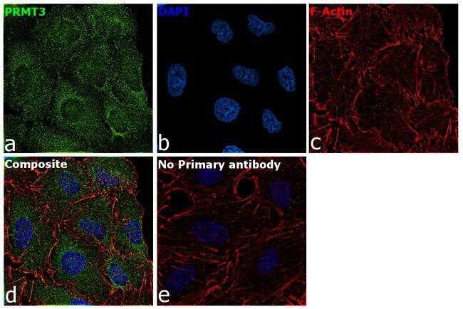
- For immunocytochemistry analysis, A549 cells were fixed and permeabilized for detection of endogenous PRMT3 using Anti-PRMT3 Recombinant Rabbit Monoclonal Antibody (Product # 703744) at a 1:100 dilution and labeled with Goat anti-Rabbit IgG (Heavy Chain) Superclonal™ Secondary Antibody, Alexa Fluor® 488 conjugate (Product # A27034) at a 1:2000 dilution. Panel a) shows representative cells that were stained for detection and localization of PRMT3 protein (green), Panel b) is stained for nuclei (blue) using ProLong™ Diamond Antifade Mountant with DAPI (Product # P36962). Panel c) represents cytoskeletal F-actin staining using Rhodamine Phalloidin (Product # R415) at a 1:300 dilution. Panel d) is a composite image of Panels a, b and c clearly demonstrating cytoplasmic localization of PRMT3. Panel e) represents control cells with no primary antibody to assess background. The images were captured at 60X magnification.
- Submitted by
- Invitrogen Antibodies (provider)
- Main image
- Experimental details
- Knockdown of PRMT3 was achieved by transfecting A549 cells with specific validated siRNA (Silencer® select Product # s19870). Immunofluorescence analysis was performed on A549 cells (untransfected, panel a-d), transfected with PRMT3 specific siRNA (panel i-l) or non-specific scrambled siRNA (panels e-h). Cells were fixed, permeabilized, and labeled with Anti-PRMT3 Recombinant Rabbit Monoclonal Antibody (Product # 703744) at a 1:100 dilution, followed by Goat anti-Rabbit IgG (Heavy Chain) Superclonal™ Secondary Antibody, Alexa Fluor® 488 conjugate (Product # A27034) at a 1:2000 dilution. Nuclei (blue) were stained using ProLong™ Diamond Antifade Mountant with DAPI (Product # P36962), and Rhodamine Phalloidin (Product # R415) at a 1:300 dilution was used for cytoskeletal F-actin (red) staining. Significant reduction of the signal was observed upon siRNA mediated knockdown (panel i-l) confirming the specificity of the antibody to PRMT3 (green). The images were captured at 60X magnification.

Supportive validation
- Submitted by
- Invitrogen Antibodies (provider)
- Main image
- Experimental details
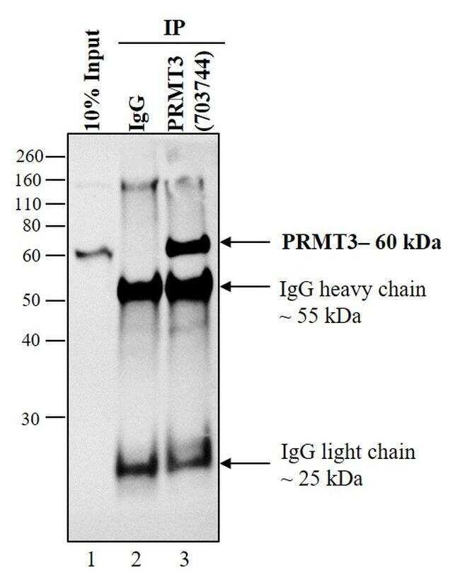
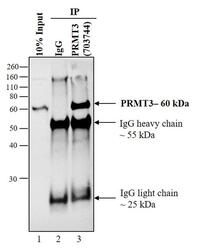
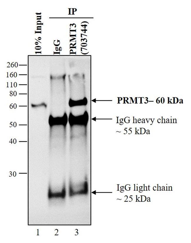
- PRMT3 was immunoprecipitated using 5 µg of the Anti-PRMT3 Recombinant Rabbit Monoclonal Antibody (Product # 703744) from whole cell extracts (800µg) of A549 (Lane 3) using the Protein A/G Dynabeads® (Product # 10001D and Product # 10003D). Normal Rabbit IgG was used as a Isotype control (Lane 2). Western blot analysis was performed using Anti-PRMT3 Recombinant Rabbit Monoclonal Antibody (Product # 703744) at a 1:5000 dilution. The blot was detected by chemiluminescence using Peroxidase IgG Fraction Monoclonal Mouse Anti-Rabbit IgG, light chain specific antibody (Product # 211-032-171) at a 1:10000 dilution. Known quantity of protein samples were electrophoresed using Novex® NuPAGE® 10% Bis-Tris gel (Product # NP03011BOX). Resolved proteins were then transferred onto a nitrocellulose membrane (Product # IB23001) by iBlot® 2 Dry Blotting System (Product # IB21001). Chemiluminescent detection was performed using Novex® ECL Chemiluminescent Substrate Reagent Kit (Product # WP20005).
Supportive validation
- Submitted by
- Invitrogen Antibodies (provider)
- Main image
- Experimental details
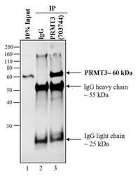
- PRMT3 was immunoprecipitated using 5 µg of the Anti-PRMT3 Recombinant Rabbit Monoclonal Antibody (Product # 703744) from whole cell extracts (800µg) of A549 (Lane 3) using the Protein A/G Dynabeads® (Product # 10001D and Product # 10003D). Normal Rabbit IgG was used as a Isotype control (Lane 2). Western blot analysis was performed using Anti-PRMT3 Recombinant Rabbit Monoclonal Antibody (Product # 703744) at a 1:5000 dilution. The blot was detected by chemiluminescence using Peroxidase IgG Fraction Monoclonal Mouse Anti-Rabbit IgG, light chain specific antibody (Product # 211-032-171) at a 1:10000 dilution. Known quantity of protein samples were electrophoresed using Novex® NuPAGE® 10% Bis-Tris gel (Product # NP03011BOX). Resolved proteins were then transferred onto a nitrocellulose membrane (Product # IB23001) by iBlot® 2 Dry Blotting System (Product # IB21001). Chemiluminescent detection was performed using Novex® ECL Chemiluminescent Substrate Reagent Kit (Product # WP20005).