 Explore
 Validate
 Learn
Learn Western blot
 Immunohistochemistry
Antibody data
- Antibody Data
- Antigen structure
- References [3]
- Comments [0]
- Validations
- Immunohistochemistry [2]
- Other assay [2]
Submit
Validation data
Reference
Comment
Report error
- Product number
- MA1-06319 - Provider product page

- Provider
- Invitrogen Antibodies
- Product name
- Cytokeratin 10 Monoclonal Antibody (RKSE60)
- Antibody type
- Monoclonal
- Antigen
- Other
- Description
- MA1-06319 detects cytokeratin 10 in human, canine, rat, porcine, zebrafish and mouse samples. MA1-06319 has sucessfully been used in Western blotting, flow cytometry, immunocytochemistry, and immunohistochemistry. The MA1-06319 immmunogens are cytokeratins from the human epidermis. Store at 4ºC or -20ºC if preferred.
- Reactivity
- Human, Mouse, Rat, Canine, Porcine, Zebrafish
- Host
- Mouse
- Isotype
- IgG
- Antibody clone number
- RKSE60
- Vial size
- 100 μg
- Concentration
- 1 mg/mL
- Storage
- Store at 4°C short term. For long term storage, store at -20°C, avoiding freeze/thaw cycles.
Submitted references Loss of FOXC1 contributes to the corneal epithelial fate switch and pathogenesis.
Core transcription regulatory circuitry orchestrates corneal epithelial homeostasis.
Epiboly generates the epidermal basal monolayer and spreads the nascent mammalian skin to enclose the embryonic body.
Li M, Zhu L, Liu J, Huang H, Guo H, Wang L, Li L, Gu S, Tan J, Zhong J, Wang B, Mao Z, Fan Y, Liu C, Yuan J, Ouyang H
Signal transduction and targeted therapy 2021 Jan 8;6(1):5
Signal transduction and targeted therapy 2021 Jan 8;6(1):5
Core transcription regulatory circuitry orchestrates corneal epithelial homeostasis.
Li M, Huang H, Li L, He C, Zhu L, Guo H, Wang L, Liu J, Wu S, Liu J, Xu T, Mao Z, Cao N, Zhang K, Lan F, Ding J, Yuan J, Liu Y, Ouyang H
Nature communications 2021 Jan 18;12(1):420
Nature communications 2021 Jan 18;12(1):420
Epiboly generates the epidermal basal monolayer and spreads the nascent mammalian skin to enclose the embryonic body.
Panousopoulou E, Hobbs C, Mason I, Green JB, Formstone CJ
Journal of cell science 2016 May 1;129(9):1915-27
Journal of cell science 2016 May 1;129(9):1915-27
No comments: Submit comment
Supportive validation
- Submitted by
- Invitrogen Antibodies (provider)
- Main image
- Experimental details
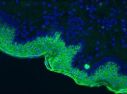
- Immunohistochemistry on frozen section of swine skin stained with Cytokeratin 10 monoclonal antibody (Product # MA1-06319). Positive staining of the keratinizing layer

- Submitted by
- Invitrogen Antibodies (provider)
- Main image

- Experimental details
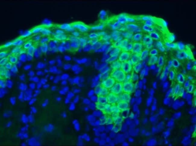
- Immunohistochemistry on frozen section of swine skin stained with Cytokeratin 10 monoclonal antibody (Product # MA1-06319). Positive staining of the keratinizing layer (higher magnification)
Supportive validation
- Submitted by
- Invitrogen Antibodies (provider)
- Main image
- Experimental details
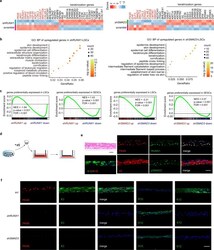
- Fig. 3 Loss of RUNX1 or SMAD3 induces cell identity switch. a Heatmaps of differentially expressed genes produced by RUNX1 or SMAD3 KD. Red mark represents SE-assigned genes. b GO BP analysis for the upregulated genes in RUNX1 -depleted and SMAD3 -depleted LSCs (pvalueCutoff = 0.01 and qvalueCutoff = 0.05). c GSEA for genes that (i) are expressed at higher levels in LSCs than in SESCs and (ii) are more highly expressed in SESCs than in LSCs. NES: normalized enrichment score. d Schema representation of the air-lifting culture system. LSCs were seeded in the transwell inserts and incubated in medium until they were confluent. Then, the medium in the upper chamber was removed to induce differentiation into a stratified epithelium sheet. e Hematoxylin and eosin (H&E) and immunofluorescence staining of the indicated genes in the differentiated corneal epithelium sheet after air-lifting induction. Scale bars, 50 mum. f Immunofluorescence staining of the indicated genes in the differentiated corneal epithelium sheets treated with the indicated shRNAs. Scale bar, 50 mum.
- Submitted by
- Invitrogen Antibodies (provider)
- Main image
- Experimental details
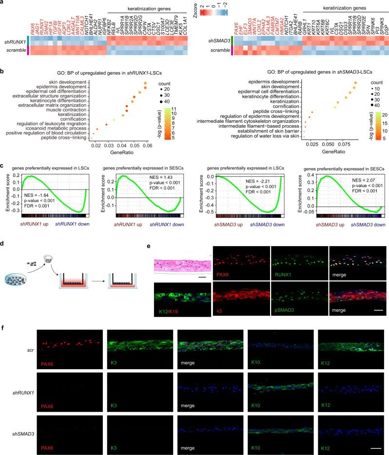
- Fig. 6 Pathological changes of corneal epithelium. H&E staining and immunofluorescence analysis of the indicated genes in corneal inflammatory (47 years old, female), ulcer (62 years old, female), alkali burn (33 years old, male), and leukoma (51 years old, female) tissues. Scale bars, 200 mum.
