Explore
 Validate
 Learn
Learn Western blot
 Immunocytochemistry
Antibody data
- Antibody Data
- Antigen structure
- References [0]
- Comments [0]
- Validations
- Immunocytochemistry [2]
- Immunohistochemistry [1]
Submit
Validation data
Reference
Comment
Report error
- Product number
- MAB4364 - Provider product page

- Provider
- R&D Systems
- Product name
- Rat Synaptotagmin-1 Antibody
- Antibody type
- Monoclonal
- Description
- Protein A or G purified from hybridoma culture supernatant. Detects Synaptotagmin-1 from rat, mouse, human, chicken, Xenopus, and fish.
- Reactivity
- Rat
- Host
- Mouse
- Conjugate
- Unconjugated
- Isotype
- IgG
- Antibody clone number
- ASV48
- Vial size
- 100 ug
- Concentration
- LYOPH
- Storage
- Use a manual defrost freezer and avoid repeated freeze-thaw cycles. 12 months from date of receipt, -20 to -70 °C as supplied. 1 month, 2 to 8 °C under sterile conditions after reconstitution. 6 months, -20 to -70 °C under sterile conditions after reconstitution.
No comments: Submit comment
Supportive validation
- Submitted by
- R&D Systems (provider)
- Main image
- Experimental details
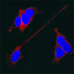
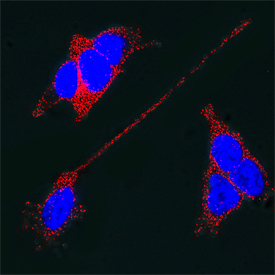
- Synaptotagmin-1 in SH-SY5Y Human Cell Line. SH-SY5Y human neuroblastoma cells were cultured overnight in the presence of 1 mM Retinoic Acid (Catalog # 0695/50) prior to immersion fixation. Synaptotagmin-1 was detected using a Mouse Anti-Rat Synaptotagmin-1 Monoclonal Antibody (Catalog # MAB4364). The cells were stained with the NorthernLights 557-conjugated Donkey Anti-Mouse IgG Affinity-purified Secondary Antibody (red; Catalog # NL007). The cell nuclei were counterstained with DAPI (blue). Synaptotagmin-1 immunoreactivity was localized to synaptic vesicles in the cytoplasm and on the plasma membrane. View our protocol for Fluorescent ICC Staining of Cells on Coverslips.
- Submitted by
- R&D Systems (provider)
- Main image

- Experimental details
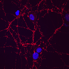
- Synaptotagmin-1 in Rat Neurons. Synaptotagmin-1 was detected in immersion fixed rat hippocampal neurons (E18) using Mouse Anti-Rat Synaptotagmin-1 Monoclonal Antibody (Catalog # MAB4364) at 10 µg/mL for 3 hours at room temperature. Cells were stained using the NorthernLights™ 557-conjugated Anti-Mouse IgG Secondary Antibody (red; Catalog # NL007) and counterstained with DAPI (blue). Specific staining was localized to synapses. View our protocol for Fluorescent ICC Staining of Cells on Coverslips.
Supportive validation
- Submitted by
- R&D Systems (provider)
- Main image

- Experimental details
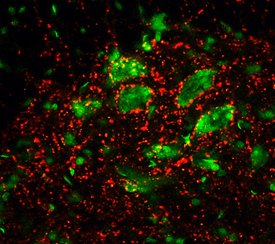
- Synaptotagmin in Rat Spinal Cord. Synaptotagmin was detected in perfusion fixed frozen sections of rat spinal cord using 8 µg/mL Mouse Anti-Rat Synaptotagmin Monoclonal Antibody (Catalog # MAB4364) overnight at 4 °C. Tissue was stained (red) and counter-stained (green). View our protocol for Fluorescent IHC Staining of Frozen Tissue Sections.