Explore
 Validate
 Learn
Learn Western blot
Antibody data
- Antibody Data
- Antigen structure
- References [0]
- Comments [0]
- Validations
- Western blot [1]
- ELISA [1]
- Immunohistochemistry [1]
Submit
Validation data
Reference
Comment
Report error
- Product number
- MA5-55665 - Provider product page

- Provider
- Invitrogen Antibodies
- Product name
- Synaptotagmin 1 Monoclonal Antibody (K1E005_2F9)
- Antibody type
- Monoclonal
- Antigen
- Recombinant full-length protein
- Description
- Sequence of this protein is as follows: KKGKEKGGKN AINMKDVKDL GKTMKDQALK DDDAETGLTD GEEKEEPKEE EKLGKLQYSL DYDFQNNQLL VGIIQAAELP ALDMGGTSDP YVKVFLLPDK KKKFETKVHR KTLNPVFNEQ FTFKVPYSEL GGKTLVMAVY DFDRFSKHDI IGEFKVPMNT VDFGHVTEEW RDLQSAEKEE QEKLGDICFS LRYVPTAGKL TVVILEAKNL KKMDVGGLSD PYVKIHLMQN GKRLKKKKTT IKKNTLNPYY NESFSFEVPF EQIQKVQVVV TVLDYDKIGK NDAIGKVFVG YNSTGAELRH WSDMLANPRR PIAQWHTLQV EEEVDAMLAV
- Reactivity
- Human, Rat
- Host
- Mouse
- Isotype
- IgG
- Antibody clone number
- K1E005_2F9
- Vial size
- 50 μg
- Concentration
- 1 mg/mL
- Storage
- Store at 4°C short term. For long term storage, store at -20°C, avoiding freeze/thaw cycles.
No comments: Submit comment
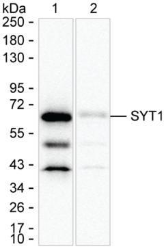
Supportive validation
- Submitted by
- Invitrogen Antibodies (provider)
- Main image
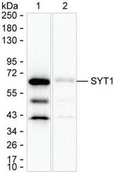
- Experimental details
- Western blot analysis of Synaptotagmin 1 in 15 µg of SH-SY5Y lysate were run on 6-18% SDS-PAGE under reducing conditions and blotted onto nitrocellulose membrane. Antibody concentration at 1 µg/mL was used as the primary antibody and peroxidase conjugated goat anti-mouse IgG was used as the secondary antibody. Synaptotagmin 1 band was visualized using ECL Substrate. Sample was run on 6-18% SDS-PAGE under reducing conditions, blotted onto nitrocellulose membrane, and peroxidase conjugated goat anti-mouse IgG was used as the secondary antibody. Synaptotagmin 1 band was visualized using ECL Substrate. Incubation with primary Synaptotagmin 1 monoclonal antibody (Product # MA5-55665) at a dilution of 1 µg/mL was used.
Supportive validation
- Submitted by
- Invitrogen Antibodies (provider)
- Main image
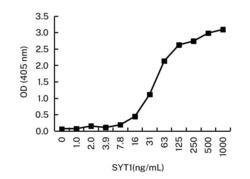
- Experimental details
- ELISA using Synaptotagmin 1 as the antigen. Microtiter wells were coated with Synaptotagmin 1 monoclonal antibody (Product # MA5-55665) at a dilution of 3 µg/mL (capture). Peroxidase conjugated mouse anti-Synaptotagmin 1 monoclonal antibody was used as the detection antibody.

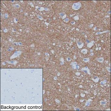
Supportive validation
- Submitted by
- Invitrogen Antibodies (provider)
- Main image

- Experimental details
- Immunohistochemistry analysis of Synaptotagmin 1 in paraffin-embedded cerebral cortex tissue. Sample was incubated with Synaptotagmin 1 monoclonal antibody (Product # MA5-55665) at a dilution of 1 µg/mL (RT, 1 hour). Antigen was retrieved through addition of boiling Tris/EDTA buffer pH 9 in a pressure cooker for 3 min. Endogenous peroxidase activity was quenched by incubating the sections with 3% H2O2 for 30 min at room temperature. Poly-peroxidase conjugated goat anti-mouse IgG was used as the secondary antibody. Diaminobenzidine was used as the chromogen. The section was counterstained with hematoxylin. A tissue section incubated with phosphate-buffered saline followed by incubation with the secondary antibody was used as the background control.